Image
Figure Caption
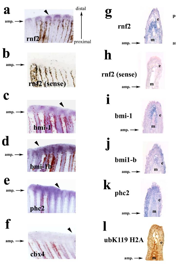
Fig. S6 Whole mount in situ hybridization of Polycomb PRC1 components during caudal fin regeneration at 48 hpa (a–f). Longitudinal sections of caudal fins demonstrating PRC1 components expression during regeneration at 48 hpa (g–k). Ubiquitous presence of Ub-H2AK119 in caudal fin detected by immunohistochemistry (l). IHC was performed on PFA fixed longitudinal sections of fins at 48 hpa. The epidermis (e) and mesenchyme (m) are indicated in each panel. Antibody signal (brown) and hematoxylin counterstain (blue) are shown.
Acknowledgments
This image is the copyrighted work of the attributed author or publisher, and
ZFIN has permission only to display this image to its users.
Additional permissions should be obtained from the applicable author or publisher of the image.
Full text @ Proc. Natl. Acad. Sci. USA

