
Fig. S3
- ID
- ZDB-IMAGE-101115-15
- Publication
- Stewart et al., 2009 - A histone demethylase is necessary for regeneration in zebrafish
- All Figures
- Figures for Stewart et al., 2009
|
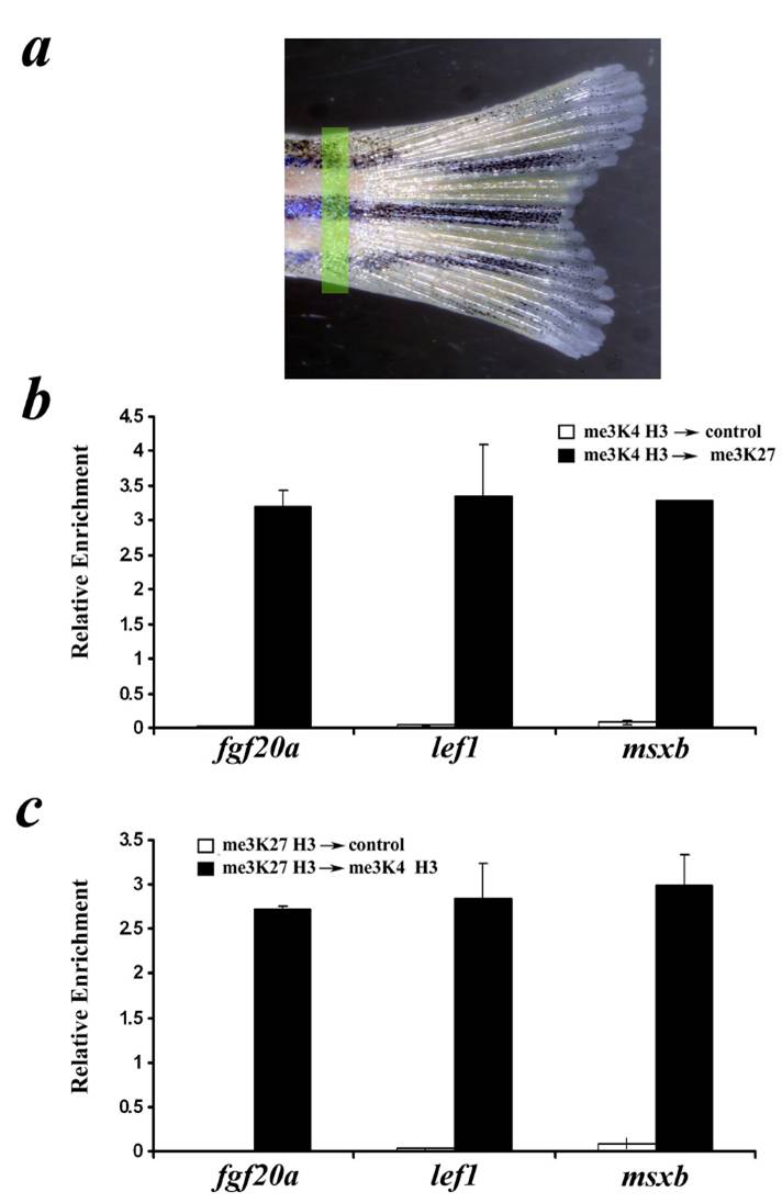
Fig. S3 (a) Zebrafish proximal caudal fin tissue. The yellow highlighted region roughly indicates the region used for chromatin extraction and ChIP analysis. (b and c) Bivalent chromatin in non-regenerating zebrafish proximal caudal fin tissue. (b) Sequential ChIP first performed with me3K4 H3 followed by control or me3K27 H3 antibody. (c) Sequential ChIP first performed with me3K27 H3 followed by control or me3K4 H3antibody. Eluates from the second ChIP were examined by qPCR using primers near the transcription start sites of the indicated genes. Shown is the average of two independent sequential ChIPs; error bars indicate deviation from the mean from two cohorts.