
Fig. 6
- ID
- ZDB-IMAGE-101111-6
- Publication
- Perälä et al., 2010 - Conservation, expression, and knockdown of zebrafish plxnb2a and plxnb2b
- All Figures
- Figures for Perälä et al., 2010
|
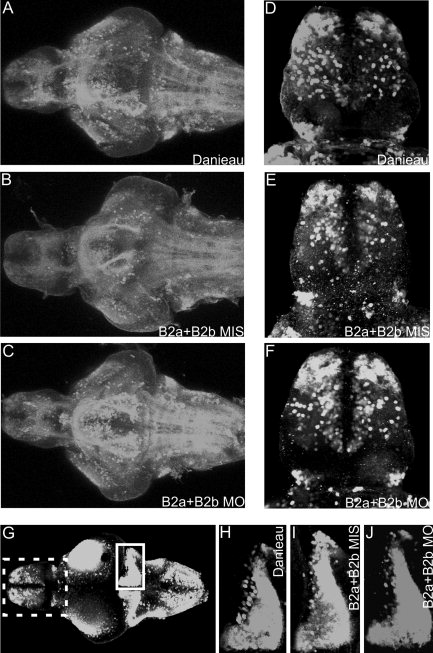
Fig. 6 The GABAergic neurons in 5 dpf zebrafish brain. A-C: A horizontal overview of stacked confocal scans from the ventral side of the brain after treatment with Danieau (A), B2aMIS+B2bMIS (5 ng+5 ng; B), or B2aMO+B2BMO (5 ng+5 ng; C). D-F: Dorsal view of the telencephalon after the injection of Danieau (D), B2aMIS+B2bMIS (E), or B2aMO+B2bMO (F) treatment, showing the GABAergic neurons in the olfactory bulb and medial telencephalon. G: Dorsal overview of the GABAergic neurons in the 5-day zebrafish brain. The dashed box (telencephalon) corresponds to the area enlarged in D-F. The cerebellar GABAergic neurons are boxed. H-J: Enlargements of the cerebellar GABAergic neuron area (boxed in G) of Danieau- (H), B2aMIS+B2bMIS- (I), and B2aMO+B2bMO- (J) treated samples.