
Fig. 3
- ID
- ZDB-IMAGE-101111-49
- Genes
- Antibodies
- Publication
- Nourizadeh-Lillabadi et al., 2010 - Early embryonic gene expression profiling of zebrafish prion protein (prp2) morphants
- All Figures
- Figures for Nourizadeh-Lillabadi et al., 2010
|
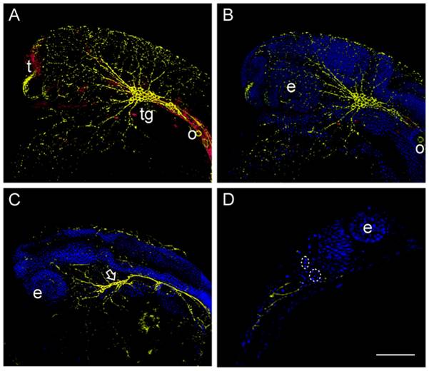
Fig. 3 Neuronal structures were visualized by the Zn12 antibody (Yellow), whereas Prp2 was detected with a rabbit anti serum (Red). A) In the wild type 24 hpf Prp2 is observed in the trigeminal ganglion and telencephalon, but also present in other neuronal tissue. B) Neuronal structures visualized by HNK-1 staining (Zn-12 antibody) in the same specimen as in A. C) Aberrant morphology of the trigeminal ganglion in Prp2 morphants (arrow) as well as reduced number of peripheral neurons visualized by HNK-1 staining. D) Non deconvolved single plane image of a morphant with condensed nuclei (inside stippled ring). Abbreviations: eye (e), telencephalon (t) and trigeminal ganglion (tg).