
Fig. 3
- ID
- ZDB-IMAGE-101111-3
- Genes
- Publication
- Perälä et al., 2010 - Conservation, expression, and knockdown of zebrafish plxnb2a and plxnb2b
- All Figures
- Figures for Perälä et al., 2010
|
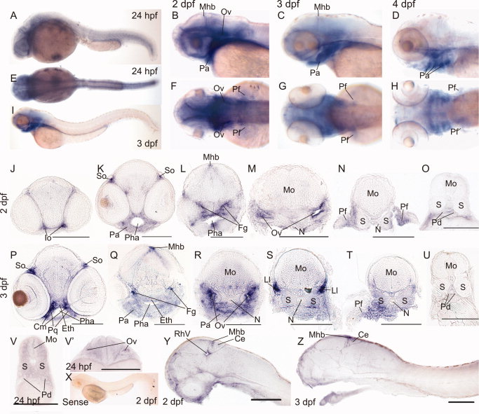
Fig. 3 Expression of plxnb2a in 24 hours postfertilization (hpf) to 4 days postfertilization (dpf) zebrafish embryos by whole-mount in situ hybridization. A-D: Lateral views. E-H: Dorsal views. I: Lateral view of a 3 dpf embryo. J-V′: Transverse sections of 2 dpf (J-O), 3 dpf (P-U), and 24 hpf (V,V′) whole-mount samples. X: No staining was seen with the control sense probe. Y,Z: Sagittal sections of 2 dpf (Y) and 3 dpf (Z) whole-mount samples. Ce, cerebellum; Cm, cranial mesenchyme; Eth, ethmoid plate; Fg, facial ganglion; Io, neuromasts of the infraorbital lateral line; Ll, lateral line ganglia; Mhb, midbrain-hindbrain boundary; Mo, medulla oblongata; N, notochord; Ov, otic vesicle; Pa, pharyngeal arches; Pd, pronephric duct; Pf, pectoral fin; Pha, pharynx; Pq, palatoquadrate; RhV, rhombencephalic ventricle; S, somite; So, neuromasts of the supraorbital lateral line. Scale bars = 100 μm in J-V′,Y-Z.