
|
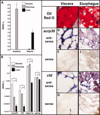
Fig. 2 Orthologs of mammalian adipocyte marker genes are enriched in adult visceral, pancreatic, and esophageal adipocytes. The liver, pancreas which contains adipocytes, and visceral white adipose tissue (WAT) were dissected from three batches of three to five adult zebrafish, pooled, and processed for quantitative polymerase chain reaction (qPCR; A,B). A:amylase is a specific marker of pancreatic acinar cells and fabp10 is only expressed in hepatocytes, demonstrating the specificity of the dissected tissues. The zebrafish orthologs of acrp30, cfd, fabp11a, and pparg are expressed specifically in visceral adipose tissue and pancreas as opposed to liver. B: Bars represent the standard deviation and *indicates P < 0.05. C–L: Oil red O staining (C,H) and in situ hybridization (D-G,I-L) was carried out on consecutive serial sections of adult fish through the visceral and esophageal WAT stores. acrp30 and cfd antisense probes show positive staining in visceral (D-F) and esophageal (I-L) adipocytes coincides with positive oil red O staining in (C,H). There is some staining with both probes in esophageal muscle adjacent to the adipocytes (I,K) which is absent from sections stained with the respective senses probe (J,L). Scale bar = 50 μm.