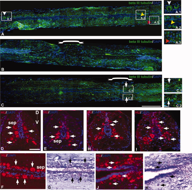
Fig. 6 Immunohistochemical staining of β-III tubulin (A-C), Hu (D–F and H-J), and counter-staining of Luxol fast blue/Cresyl violet (G and K) in zebrafish uninjured (A, D–G) and injured spinal cord (B,C, and H–K). A: Section of uninjured cord shows the spatial distribution of different types of neurons sensory neurons (white ▼), interneurons close to ependyma (yellow ▼), and motorneurons (red ▼). A.1, A.2, A.3: Higher magnification of indicated areas of A showing different types of neurons. B: 3dpi cord section showing the loss of mature neurons at the injury epicenter. C: One month post-injured cord section showing regenerated neurons at the injury epicenter. C.1, C.2: Higher magnification of indicated areas in C, where formation of new neurons (thick arrow) both at the dorsal and ventral sides can be seen. D–F: Sections of uninjured cord showing the distribution of Hu+ neurons (thick arrow) in the subependyma (sep) along the D-V axis. G: Luxol fast blue/Cresyl violet staining of F. H–J: Sections from 10dpi cord showing the presence of Hu+ newly formed neurons (thick arrow) in different locations along the D-V axis. K: Staining of the same section in J with Luxol fast blue/Cresyl violet. Scale bar = 250 μm (A-C), 50 μm (A.1-A.3, C.1,C.2, D-K).
Image
Figure Caption
Acknowledgments
This image is the copyrighted work of the attributed author or publisher, and
ZFIN has permission only to display this image to its users.
Additional permissions should be obtained from the applicable author or publisher of the image.
Full text @ Dev. Dyn.

