
Fig. 4
- ID
- ZDB-IMAGE-101021-44
- Genes
- Publication
- Appelbaum et al., 2010 - Circadian and homeostatic regulation of structural synaptic plasticity in hypocretin neurons
- All Figures
- Figures for Appelbaum et al., 2010
|
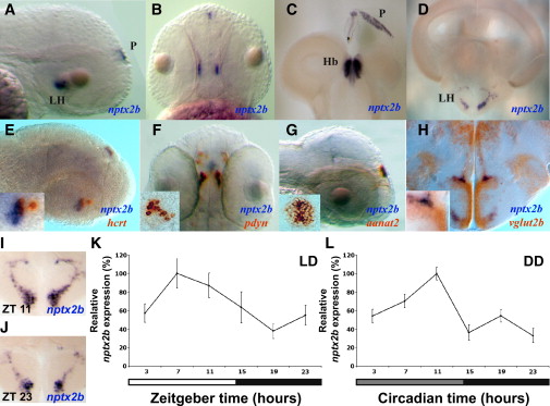
Fig. 4 Nptx2b Cell Identity and Rhythmic nptx2b Expression in the Lateral Hypothalamus
(A and B) Lateral and dorsal views of 2 dpf larvae expressing nptx2b in the lateral hypothalamus (LH) and the pineal gland (P).
(C and D) Similar pattern of nptx2b expression is shown in the LH (D), the habenula (Hb, C), and the pineal gland (C) in adult fish.
(E–G) Double ISH in 2 dpf larvae showing colocalization of nptx2b with hcrt (E) and pdyn (F) in the LH and with aanat2 in the pineal gland (G).
(H) Double ISH in adult shows that nptx2b neurons express vglut2b, a marker of glutamatergic neurons.
(I–L) Time-course analysis under light:dark (LD) and constant darkness (DD, gray bar represents subjective day) conditions demonstrates that endogenous nptx2b expression increases during the day and decreases during the night (K and L, n = 7 adult brains per time point, p < 0.001). Representative adult brain sections for day (I) and night (J) are shown. Statistical comparisons were performed with ANOVAs. Each value represents mean ± SEM.