
Fig. 6
- ID
- ZDB-IMAGE-101013-2
- Antibodies
- Publication
- Van Otterloo et al., 2010 - Differentiation of zebrafish melanophores depends on transcription factors AP2 alpha and AP2 epsilon
- All Figures
- Figures for Van Otterloo et al., 2010
|
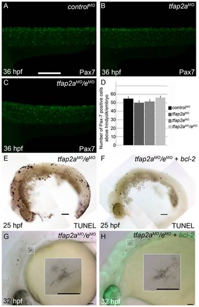
Fig. 6 Contribution of cell fate specification and cell death to melanophore defects in tfap2a/e doubly-deficient embryos.
(A–C) Lateral views of 36 hpf embryos stained with α-Pax7 to mark xanthophores. A similar number of α-Pax-7 IR positive cells is apparent in wild-type embryos injected with (A) a control MO, (B) the tfap2a MO, and (C) the tfap2a MO/tfap2e MO. (D) Average values for the number of α-Pax-7 IR positive cells counted above the hind yolk, n = 10 embryos per group. (E, F) Lateral views of 25 hpf embryos processed with the TUNEL reaction. (E) In an embryo injected with tfap2a/e MO alone there are many more TUNEL-positive cells than in (F) an embryo co-injected with an mRNA encoding a bcl2-gfp mRNA. (This effect was quantified in a parallel experiment, in which bcl2GFP mRNA was co-injected with control MO, embryos fixed at 24 hpf, and the number of TUNEL-positive cells counted: control MO, 97.7±15.5; control MO + bcl2-gfp 54.4±12.3, Avg±SE, p = 0.03). (G–H) Lateral views of live 32 hpf embryos. (G) In an embryo injected with the tfap2a/e MO alone, or (H) in an embryo co-injected with bcl2-gfp mRNA, melanophores appeared similarly poorly differentiated. Insets in G and H, higher magnification views of melanophores in the respective embryos. Scale bars: (A–C, E–H) 100 μM; (Insets in G–H) 50 μM.