
|
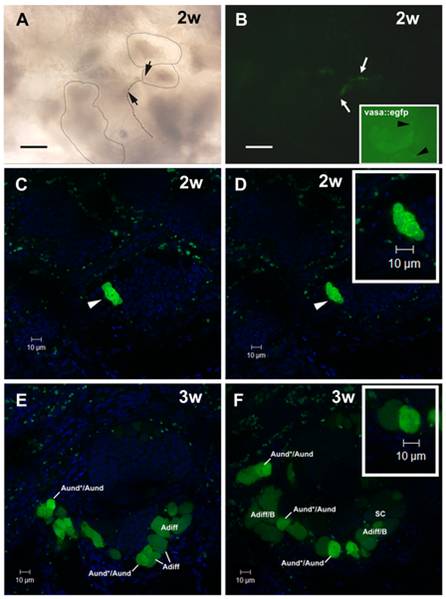
Fig. 5 SSC transplantation into male zebrafish recipients.
A,B. Recipient testes after two weeks (2w) of transplantation analyzed by light (A) and fluorescence (B) microscopies. Seminiferous tubules are delimited by stippled lines. Arrows indicate the same area in A and B. Donor cells formed clusters situated near the interstitium in a similar way as observed in vasa::egfp testes (see arrowheads in the inset). Nuclei (blue) are stained with DAPI. Scale bars = 50 μm. C,D. CLSM analysis of recipient testes after two weeks (2w) of transplantation. Arrowheads indicate a donor-derived cyst composed of ~8 cells. Inset. High magnification of donor-derived cyst. Nuclei (blue) are stained with DAPI. E,F. Recipient testes after 3 weeks (3w) of transplantation analyzed under CLSM. Donor-derived cysts increased their number and size, being found at different stages of spermatogenesis. Type A undifferentiated spermatogonia (Aund*/Aund), type A differentiating spermatogonia (Adiff), type B spermatogonia (B), and spermatocytes (SC). Inset shows a high magnification of type A undifferentiated spermatogonia. Nuclei (blue) are stained with DAPI.