
Fig. 4
- ID
- ZDB-IMAGE-101004-22
- Genes
- Publication
- Whitfield et al., 1996 - Mutations affecting development of the zebrafish inner ear and lateral line
- All Figures
- Figures for Whitfield et al., 1996
|
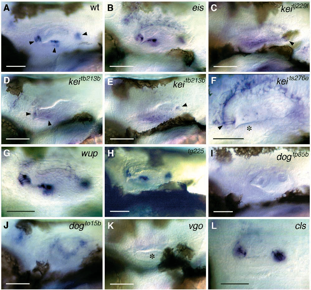
Fig. 4 mshC expression in ears of the mutants. In situ hybridisations to mshC mRNA in ears at 48 hours of development. Gene names are given for each panel; see text for details. Lateral views; anterior to the left. (A) Wild type (wt). Note the strong expression in three patches (anterior, lateral and posterior; arrowheads), thought to represent the developing cristae. The maculae do not appear to express mshC. (C-E) Expression in kei embryos is delocalised. D and E are views of the same ear at different focal levels, to show limited localisation to three patches (arrowheads) in addition to delocalised staining. (F) Close up of kei anterior patch of expression. The asterisk marks the developing anterior macula, which does not express mshC. (I,J) dog: in I, the allele dogtp85b does not show any expression, while in dogto15b, delocalised and weak expression is seen. (K) vgo: no expression is evident. Asterisk marks the single macula, with nuclei in two distinct layers, as in the wild type. L, cls: only two patches of mshC expression are evident. Scale bars, 50 μm.