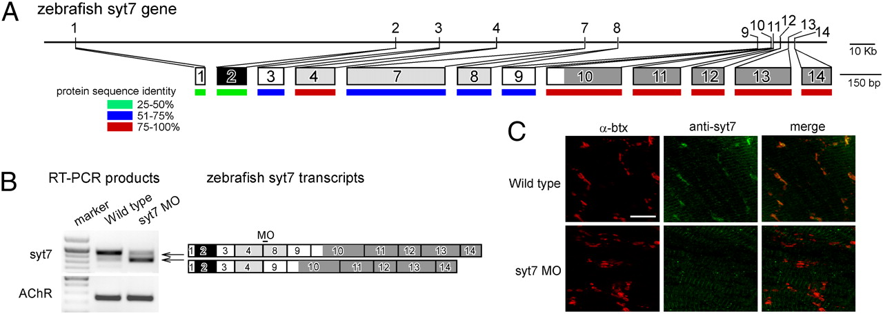
Fig. 4 Zebrafish syt7 gene and transcripts. (A) The location of exons on the gene (Upper) and transcript (Lower) are shown for zebrafish syt7. Exons are numbered on the basis of the mammalian gene (25, 26). The relative sizes of individual exons are indicated, along with the percentage identity to rat syt7 on the amino acid level (color indicators). The domains, predicted on the basis of homology to the mammalian gene are: transmembrane (black), calcium-binding C2 domains (dark gray), and alternatively spliced (light gray) regions. (B) RT-PCR analysis on wild-type and syt7 morpholino fish. The exon structures of the RT-PCR products and morpholino position are shown schematically. As a control the α-subunit of muscle acetycholine receptors was amplified from the same sample. (C) Syt7 antibody label (green) colocalizes with α-btx labeling (red) in wild type (Upper) but is absent at α-btx-labeled sites in syt7 morpholino fish (Lower). (Scale bar, 10 μm.)
Image
Figure Caption
Figure Data
Acknowledgments
This image is the copyrighted work of the attributed author or publisher, and
ZFIN has permission only to display this image to its users.
Additional permissions should be obtained from the applicable author or publisher of the image.
Full text @ Proc. Natl. Acad. Sci. USA

