
Fig. 4
- ID
- ZDB-IMAGE-100903-53
- Genes
- Antibodies
- Publication
- Wang et al., 2010 - Moesin1 and Ve-cadherin are required in endothelial cells during in vivo tubulogenesis
- All Figures
- Figures for Wang et al., 2010
|
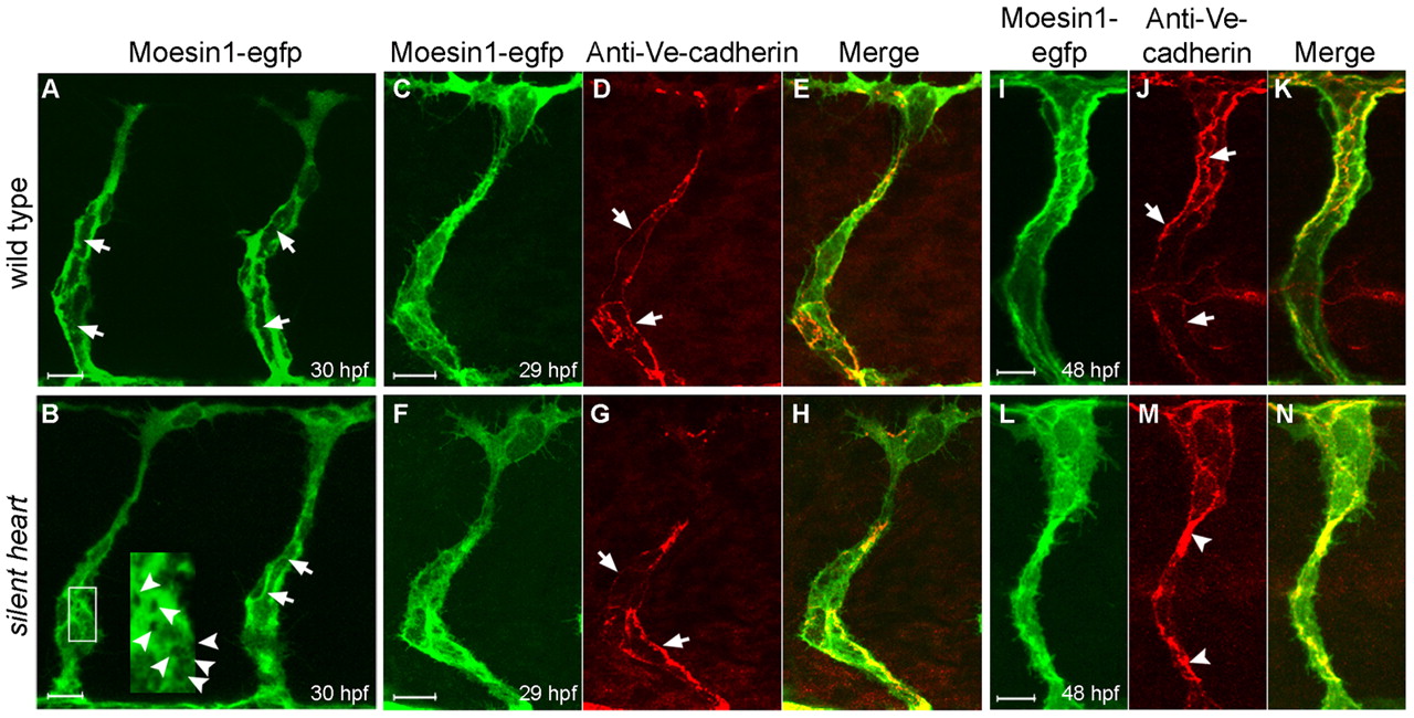
Fig. 4 Primary endothelial lumen formation in the ISVs does not require blood flow. (A,B) Formation of the primary lumen (arrows) at 30 hpf is observed with the Tg(flk1:moesin1-egfp) line in either wild-type (A) or silent heart mutants (B). (B) silent heart embryos also displayed vacuoles during tubulogenesis (arrowheads in inset). (C-H) Ve-cadherin-labeled junctions (arrows) appeared normal at 29 hpf in silent heart mutants. (I-N) At 48 hpf, Ve-cadherin-labeled junctions (arrows) are often clustered in the silent heart embryos (arrowheads), which is likely to reflect the collapse of the primary lumen.