
|
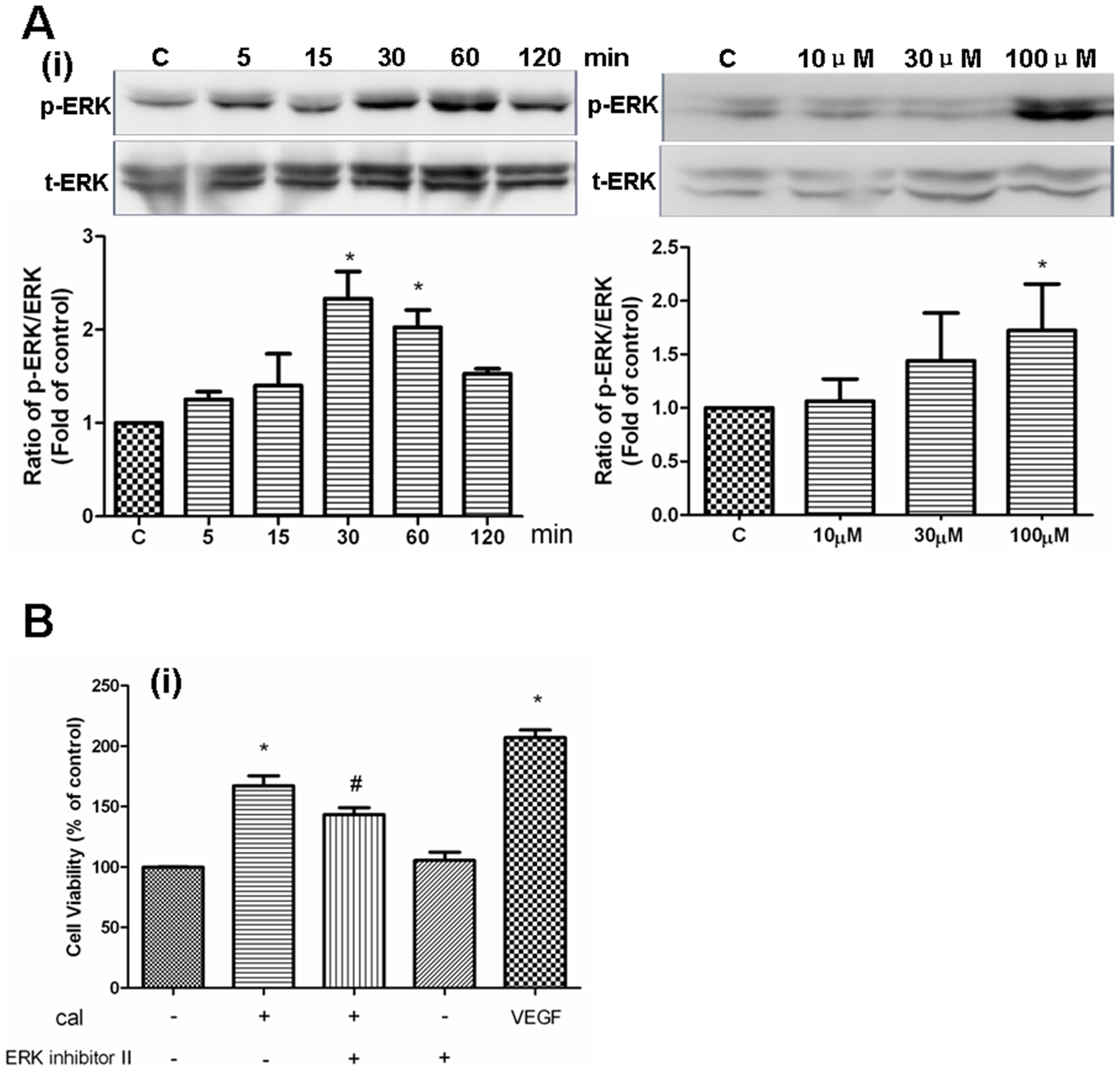
Fig. 8 Role of MAPK signaling in calycosin-induced angiogenesis.
(A) Effects of calycosin on ERK1/2 activation. HUVEC were incubated with calycosin (100 μM) at indicated time or with calycosin in different concentrations for 30 min. Expressions of phospho-ERK1/2 and total-ERK1/2 were analyzed by western blotting and quantified by densitometry. The values indicate the relative densitometric units. Results are represented as mean±SEM (n = 3 independent experiments), * P<0.05 versus control. (B) Effect of ERK activation inhibitor peptide II on calycosin-induced HUVEC proliferation. HUVECs were pre-treated with 0.5 μM ERK activation inhibitor peptide II (ERK inhibitor II) for 1 h before the addition of calycosin (100 μM). Changes in HUVEC proliferation were determined 48 h later by XTT assay. 20 ng/ml VEGF was used as the positive control in this experiment. “cal” is the abbreviation of calycosin. Results are expressed as percentage of vehicle control (100%) in mean±SEM (n≥3 independent experiments), *P<0.05 versus vehicle control, # P<0.05 versus calycosin.