
|
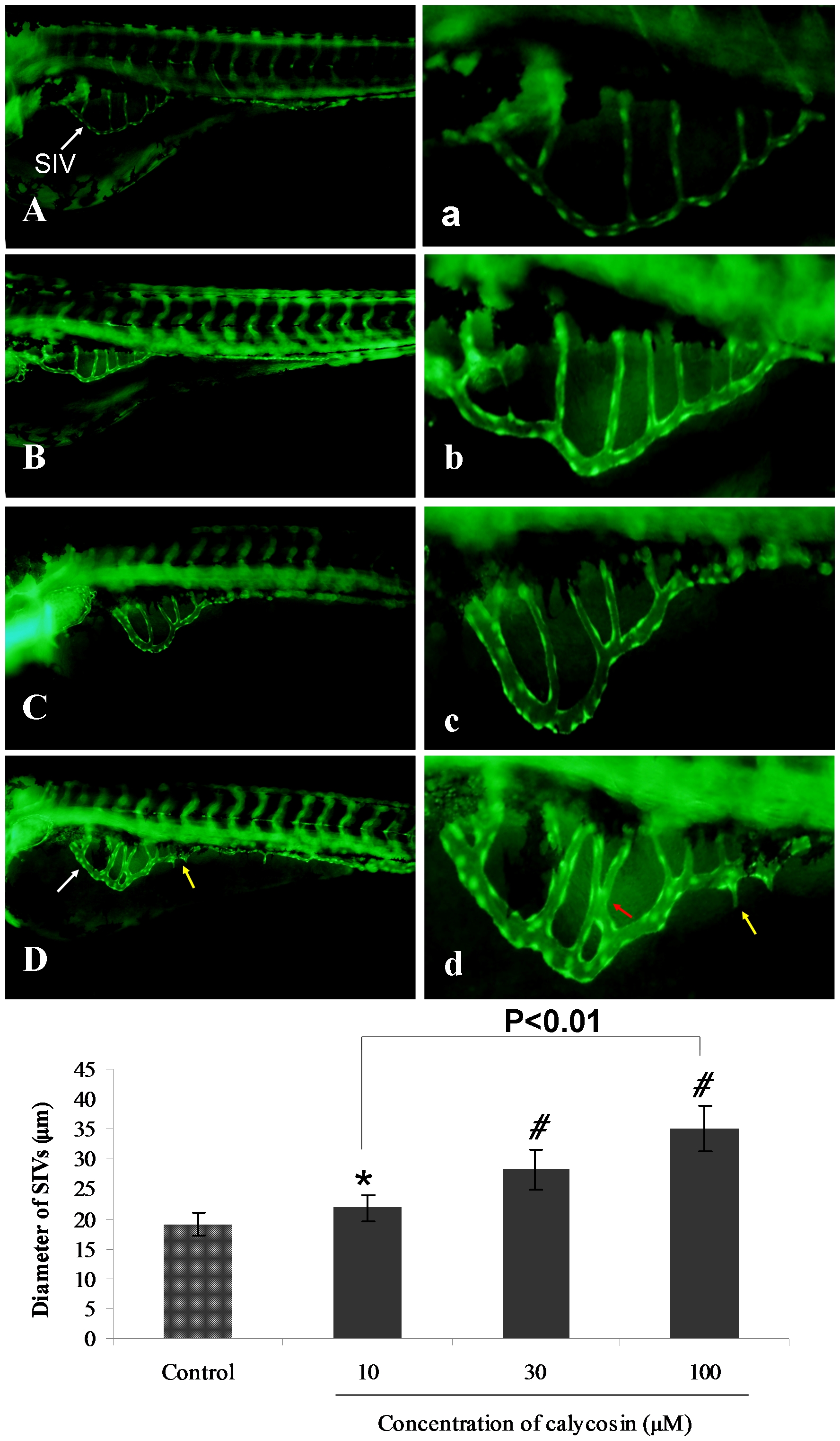
Fig. 1 The effects of calycosin treatment on blood vessel formation in SIVs of Tg(fli1:EGFP) zebrafish embryos.
(A) Control: embryo treated with 0.1% DMSO at 96 hpf, SIVs appear as a smooth basket-like structure. (B–D) Calycosin: embryo treated with 10, 30, 100 μM calycosin at 72 hpf for 24 h, leads to enlarged SIV basket stretching into the posterior yolk extension. (a–d) Enlarged SIV region (x4.5) of A–D respectively. White arrows indicating the enlarged vessels, yellow and red arrows indicate sprouting and intersectioning branches respectively. (E) Calycosin increases SIV diameter in a dose-dependent manner. Data are plotted as mean±SEM, (n = 3), *P<0.05, #P<0.001.