
|
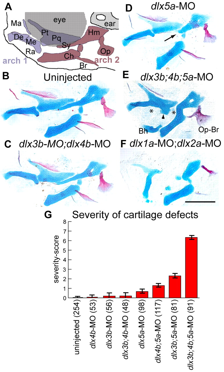
Fig. 1 Dlx function is required in intermediate domain skeleton. (A) Schematic of facial skeleton. Anterior is towards the left, dorsal is upwards. (B-F) Alcian Blue (cartilage) and Alizarin Red (bone) stained pharyngeal skeletons with Dlx morpholino treatments at 6 dpf. (B) Uninjected, (C) dlx3b-MO;dlx4b-MO and (D) dlx5a-MO fish look very similar, although dlx5a-MO sometimes causes shortened symplectic cartilages (arrow). (E) Injection of dlx3b;4b;5a-MO frequently causes dramatic skeletal defects, including joint loss (asterisks), fusion of OP and BSR bones (Op-Br), and ectopic processes attached to the palatoquadrate (arrowhead), or ventrally in the face. (F) By contrast, dlx1a-MO;dlx2a-MO injection causes defects in both dorsal and intermediate cartilages. (G) Plot of severity scores, showing that dlx3b-MO, dlx4b-MO and dlx5a-MO interact to create more than additive changes in intermediate domain skeletal phenotypes. Error bars are 95% confidence intervals, determined by ANOVA. Fish were scored bilaterally for prominent cartilage defects: first arch joint fusions, second arch joint fusions, symplectic defects, palatoquadrate defects and ectopic cartilages. Although each phenotype was seen at a range of expressivity, we assigned any defect a score of `1′, irrespective of expressivity. The `severity-score′ is the sum of these defects for both sides of the fish. Skeletal elements indicated in A are the first arch-derived Meckel′s cartilage (Me), including its retroarticular process (Ra), palatoquadrate (Pq) cartilage and its pterygoid process (Pt), as well as maxillary (Ma) and dentary (De) bones. The second arch gives rise to the ceratohyal cartilage (Ch), the hyosymplectic cartilage, which comprise distinctive hyomandibular (Hm) and symplectic (Sy) regions, as well as opercle (Op) and branchiostegal (Br) bones. A remnant of the basihyal cartilage (Bh) remains attached to the Ch in (E), as a mounting artifact. Scale bar: 100 μm.