
|
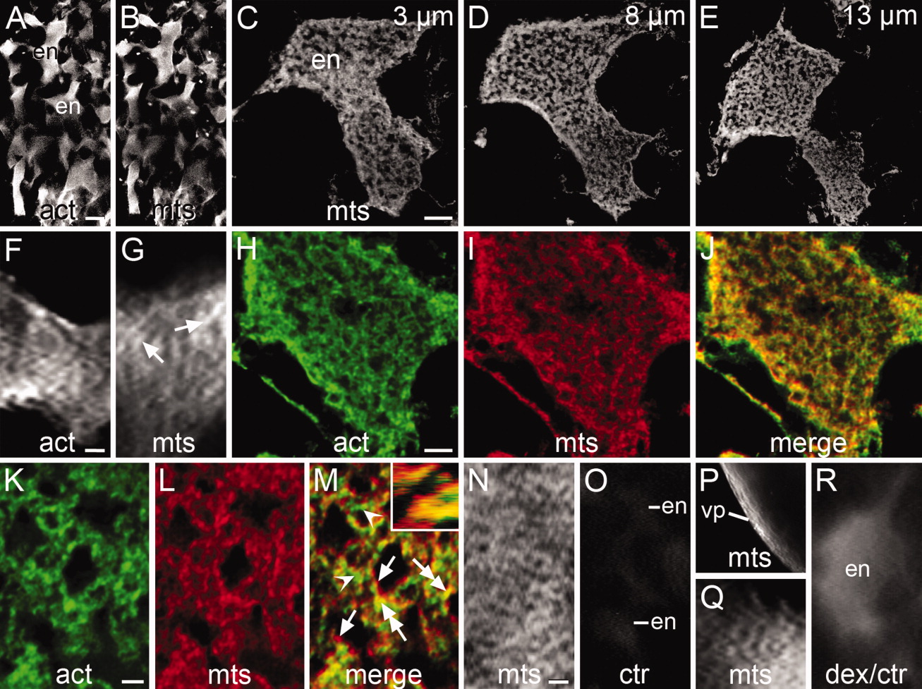
Fig. 4 Organization of the cytoskeleton in whole-mounted early stage 1b zygotes immunostained for actin filaments (act) and/or microtubules (mts) viewed under the confocal (A-E, H-J, K-M) or fluorescent (F, G, N-R) microscope. A,B: Confocal low-magnification images of the endoplasmic lacunae (en) stained for actin filaments (act) and microtubules (mts). C-E: Deconvolved confocal images selected from a z-stack containing 15x 1 μm-thick optical sections across an endoplasmic lacuna stained for microtubules. The position of the section within the stack is indicated. F: Lacuna showing a disorganized network of actin filaments. G: Lacuna showing dispersed bundles of microtubules (arrows). H-J: Deconvolved confocal intermediate magnification images of double immunostained lacuna selected from a z-stack of 15x 1-μm-thick optical sections. Shown is the network of actin filaments (H) and microtubule bundles (I) and the merged image of them (J). In the latter image, cytoskeletal bundles are seen as green dots (actin), red dots (microtubules), or yellow to orange dots (mixed actin and microtubules). K-M: Deconvolved confocal high-magnification images of double-stained lacuna, taken from a z-stack of 18x 1-μm-thick optical sections. It shows a loosely arranged cytoskeletal network built upon intercalated bundles of actin filaments (arrowheads), bundles of microtubules (arrows), and mixed bundles of actin filaments and microtubules (double-headed arrows). This condition is also illustrated in the optical cut along the X axis shown in the inset of M. N: Network of microtubules in the ectoplasm. O: Control incubated in the second antibody only. Lacunae are barely seen after a 20-second exposure. P: Bundles of oriented microtubules in the ectoplasm of the vegetal pole (vp). Q: Weakly stained microtubules in a lacuna immunostained according to Jesuthasan and Strähle ([1996]). R: Control showing an endoplasmic lacuna filled with rhodamine-labeled dextran. Notice the absence of fibrilar profiles. Scale bars = 10 μm (A-E), 6 μm (F), 5.5 μm (G), 5 μm (H-J), 2 μm (K-M), 5.6 μm (N), 10.5 μm (O), 15 μm (P), 4 μm (Q), 2 μm (R).