
|
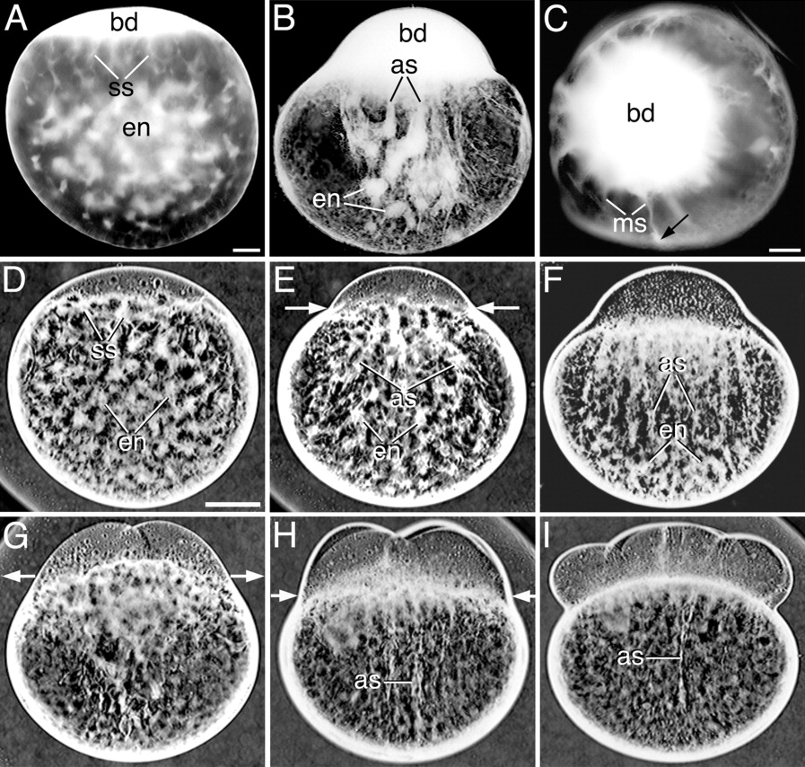
Fig. 3 Light microscope images showing the reorganization of the endoplasm (en) in inverted contrast acid-fixed (A-C) and live whole-mounted zygotes and embryos (D-I) during the first 3 cell cycles. A: Early first interphase, lateral view. B: Early first cleavage division, lateral view. C: Early first cleavage division, animal pole view. The arrow in C points to the peripheral localization of a meridional streamer (ms). Lateral views of: D: Early first interphase. E,F: Early first cleavage division. G: Two-cell embryo. H: Four-cell embryo. I: Eight-cell embryo. as, long axial streamers; bd, blastodisc; ss, short streamers. Scale bars = 80 μm (A-C), 160 μm (D-I).