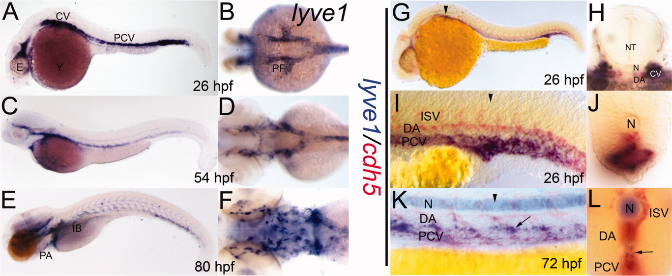
Fig. 2 A-F: lyve1 riboprobe labeled developing lymphatic endothelial cells in 26 (A,B), 54 (C,D), and 80 (E,F) hours postfertilization (hpf) embryos (A,C,E, lateral view; B,D,F, dorsal view of anterior aspect). G-L: Double staining with lyve1 (blue) and cdh5 (red) riboprobes. G-J at 26 hpf; K,L at 72 hpf; G,I,K lateral views; H,J,L transverse sections approximately at the level of black arrowheads. K,L: The lyve1 expression within the developing thoracic duct (arrows). Background label within the notochord (N) due to extended staining time. CV, cardinal vein; DA, dorsal aorta; E, eye; IB, intestinal bulb; ISV, intersegmental vessels, NT, neural tube; PF, pectoral fin bud; PA, pharyngeal arches; PCV, posterior cardinal vein; Y, yolk.
Image
Figure Caption
Figure Data
Acknowledgments
This image is the copyrighted work of the attributed author or publisher, and
ZFIN has permission only to display this image to its users.
Additional permissions should be obtained from the applicable author or publisher of the image.
Full text @ Dev. Dyn.

