
Fig. 5
- ID
- ZDB-IMAGE-100706-30
- Genes
- Publication
- Pujol-Martí et al., 2010 - Progressive neurogenesis defines lateralis somatotopy
- All Figures
- Figures for Pujol-Martí et al., 2010
|
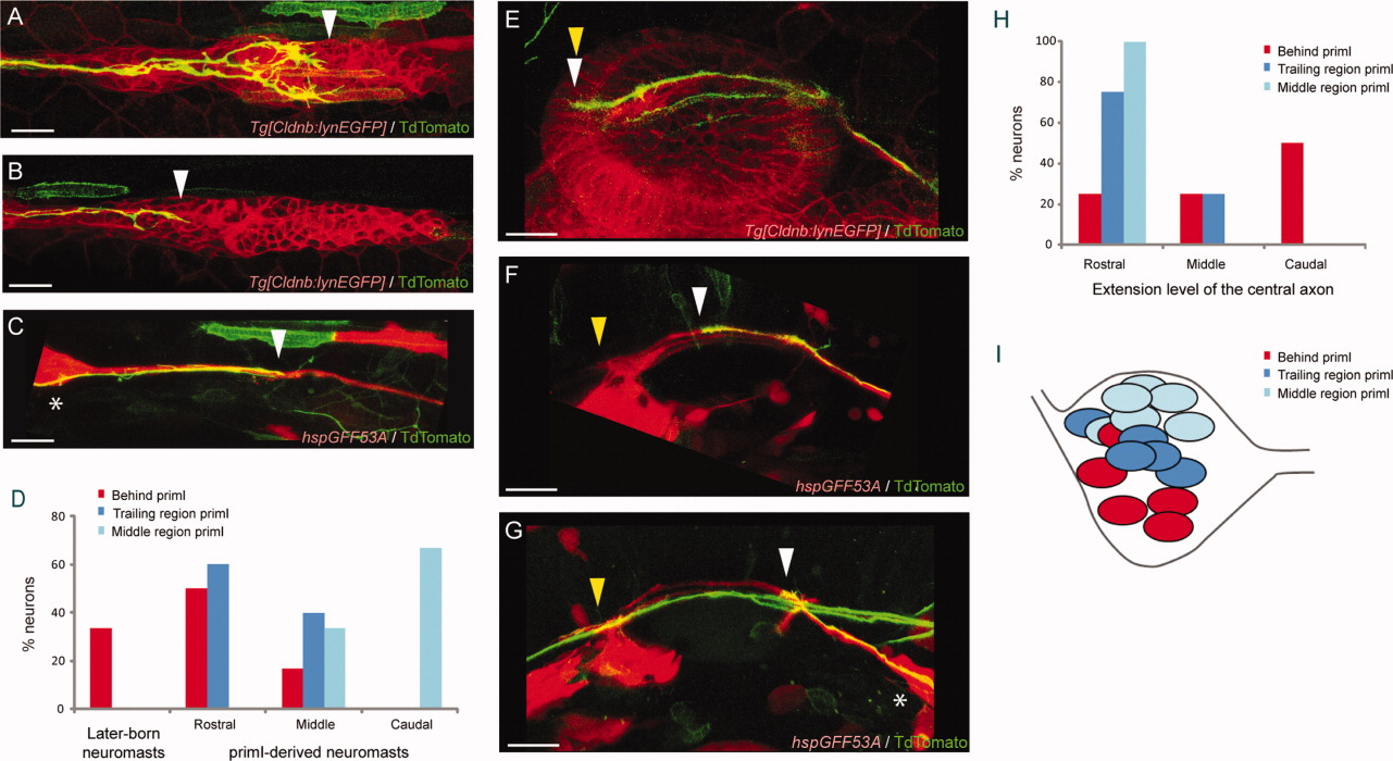
Fig. 5 Analysis of single lateralis afferent neurons. A-C, E-G: Single neurons labeled with mem-TdTomato. Eggs of Tg[CldnB:lynEGFP] or hspGFF53A;UAS:EGFP transgenic animals (red) were injected with HuC:mem-TdTomato (green). A-C: Peripheral projections of single labeled neurons at ∼27 hpf. White arrowheads indicate the position of the growth cone: middle region of primI (A), trailing region of primI (B), or behind primI (C). EGFP fluorescent signal shows the moving posterior primordium in A and B. Growth cones that do not touch the primordium are not arborized (C). Asterisk in C indicates the posterior lateralis ganglion. D: Percentage of neurons with growth cones in the middle region of primI (light blue), trailing region of primI (dark blue), or behind primI (red), which show different neuromast selection at 3.5 dpf. E-G: Central projections of single labeled neurons at ∼27 hpf. White arrowheads indicate the position of the hindbrain reached by the mem-TdTomato-labeled neurons: rostral (E), middle (F), or caudal (G). Yellow arrowheads indicate the position of the hindbrain reached by the EGFP(+) neurons. Asterisk in G indicates the posterior lateralis ganglion. H: Percentage of neurons with growth cones in the middle region primI, trailing region primI, or behind primI, which show a different extension of the central projection at ∼27 hpf. I: Somata location in the posterior lateralis ganglion at 3.5 dpf of mem-TdTomato-labeled neurons of the 3 types: growth cones in the middle region primI, trailing region primI, or behind primI. All images are maximal projections and lateral views. Dorsal is towards top, anterior is towards left. Scale bars = 30 μm (E-G), 20 μm (A-C).