
Fig. 5
- ID
- ZDB-IMAGE-100628-5
- Genes
- Publication
- Cheah et al., 2010 - tgfbeta3 Regulation of Chondrogenesis and Osteogenesis in Zebrafish is Mediated Through Formation and Survival of a Subpopulation of the Cranial Neural Crest
- All Figures
- Figures for Cheah et al., 2010
|
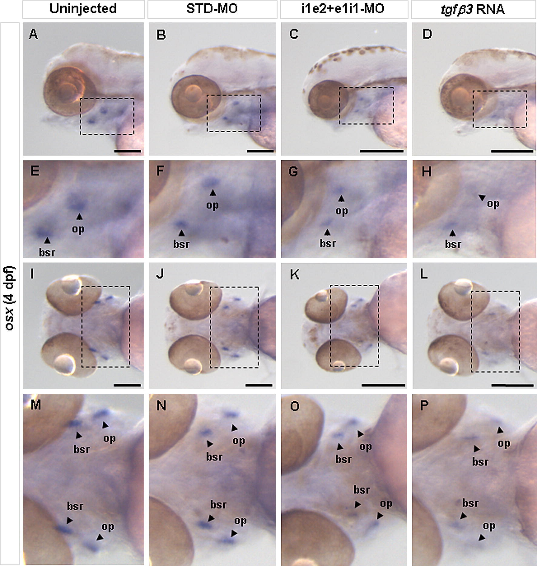
Fig. 5 Effect of tgfβ3 knockdown and over-expression on osteoblast differentiation in 4 dpf larvae. Whole-mount lateral (A–D) and ventral (I–L) views are shown. E–H and M–P show the zoom-in view of the dashed box area in A–D and I–L, respectively. Analysis of the osteoblast differentiation marker osx in 4 dpf hatchlings reveals that, compared to uninjected (A, E, I, M) and STD-MO injected (B, F, J, N) hatchlings, both the double knockdown morphant (C, G, K, O) and tgfβ3 over-expressing hatchling (D, H, L, P) show markedly reduced expression in the pharyngeal arch region, most notably in the opercle (op) and branchiostegal ray (bsr). Black horizontal scale bars represent 200 μm.
Reprinted from Mechanisms of Development, 128(7-8), Cheah, F.S., Winkler, C., Jabs, E.W., and Chong, S.S., tgfbeta3 Regulation of Chondrogenesis and Osteogenesis in Zebrafish is Mediated Through Formation and Survival of a Subpopulation of the Cranial Neural Crest, 329-344, Copyright (2010) with permission from Elsevier. Full text @ Mech. Dev.