
|
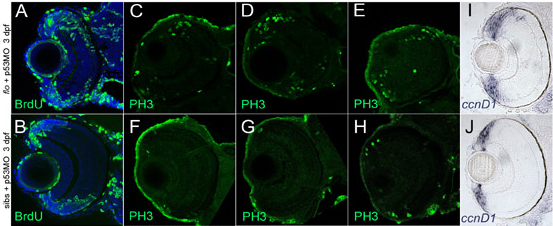
Fig. S5 flo retinae exhibit increased proliferation in/near the CMZ. (A-H) Markers for the S (BrdU) and M (PH3) phases of the cell cycle are elevated in flo + p53MO embryos at 3 dpf. Frontal transverse cryosections stained with anti-BrdU (A,B, green) or anti-PH3 (C-H, green) to identify proliferating (A,B) or mitotic (C-H) cells. Nuclei in A,B are counterstained with To-Pro 633 (blue). (C-H) Of the sections examined (n=10 eyes, three representative examples of each are shown), WT retinae contained 2-8 PH3+ cells near the periphery, whereas the number of PH3+ cells in flo retinae ranged from 16-32. (I,J) Additionally, the region of the CMZ marked by cyclin D1 expression (ccnd1) is noticeably expanded when apoptosis is blocked in flo embryos.