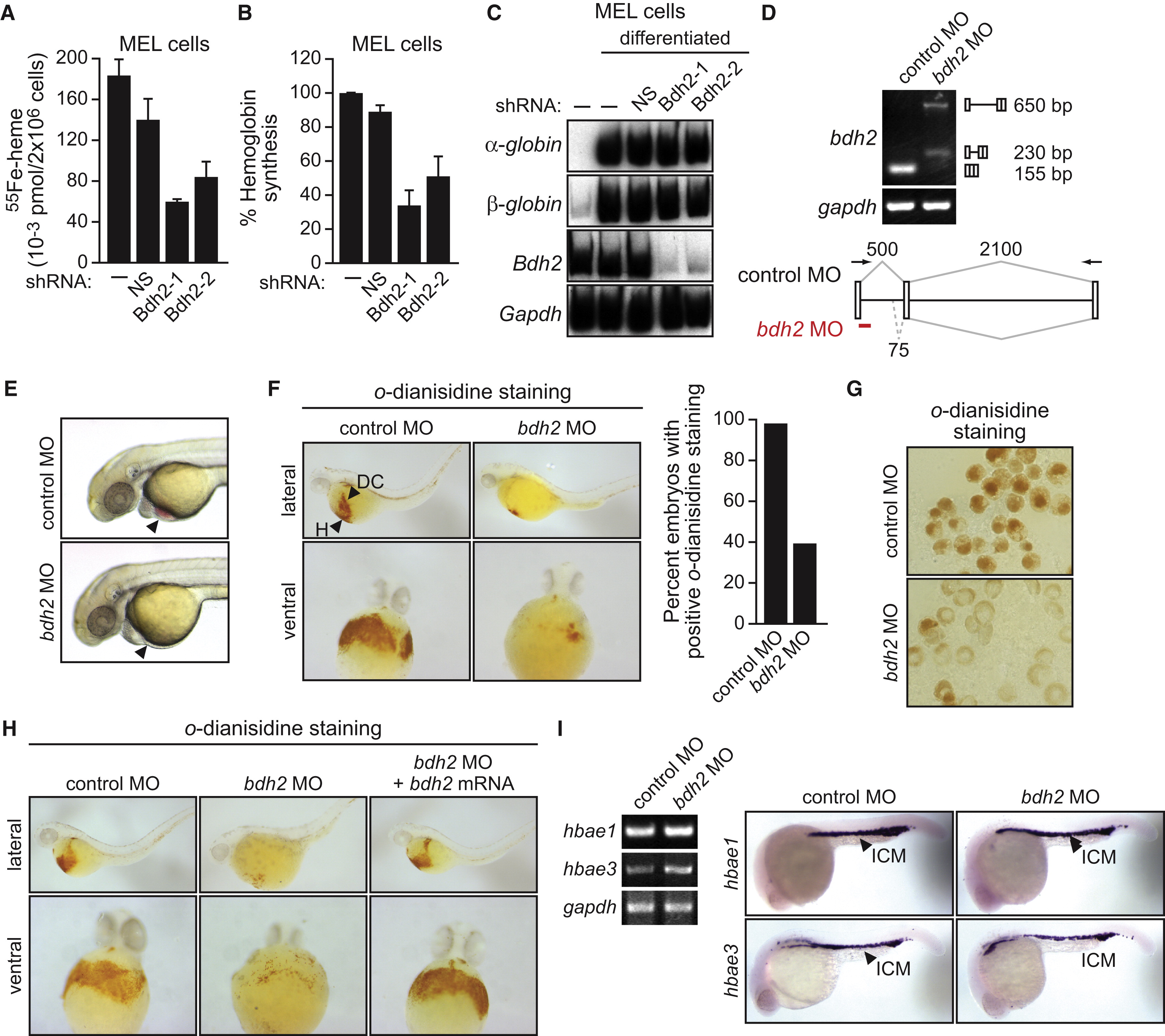
Fig. 7 Requirement of Vertebrate EntA Homologs for Heme Synthesis
(A) MEL cells expressing a NS or Bdh2 shRNA were differentiated. After labeling with 55Fe, cells were lysed, and heme was extracted and analyzed for 55Fe incorporation. Error bars indicate the SD.
(B) MEL cells were differentiated as described in (A), and after cell lysis, hemoglobin levels were determined by benzidine reaction. Hemoglobin content was expressed relative to that in untreated MEL cells cultured in the absence of dimethyl sulfoxide. Error bars indicate the SD.
(C) MEL cells were differentiated as described in (A), and α- and β-globin mRNA levels were monitored by RT-PCR. The levels of Bdh2 and Gapdh were monitored as controls.
(D) RT-PCR analysis. The bdh2 MO was injected into one-cell-stage embryos, and 24 hr later, RNA was extracted and analyzed by RT-PCR. Treatment with the bdh2 MO resulted in retention of the entire intron (upper band) as well as activation of a cryptic splice site that resulted in partial intron retention (lower band).
(E) Phenotypic analysis of control MO- and bdh2 MO-injected embryos at 72 hr postfertilization (hpf). Bdh2-depleted embryos lack hemoglobinized erythrocytes, as evidenced by the absence of red color (arrow).
(F) Whole-mount o-dianisidine staining of control MO and bdh2 MO-injected embryos at 72 hpf. Shown are lateral (top) and ventral (bottom) views of the anterior region of embryos. Control MO-injected embryos show positive o-dianisidine staining at the duct of Cuvier (DC) and the heart (H), which is markedly reduced in bdh2 MO-injected embryos. Right: quantification of the percent embryos with normal o-dianisidine staining.
(G) Blood cells were isolated from control or bdh2 MO-injected embryos and analyzed for hemoglobinization by o-dianisidine staining.
(H) Whole-mount o-dianisidine staining of embryos (72 hpf) injected with a control MO or bdh2 MO, or bdh2 MO-treated embryos coinjected with a bdh2 mRNA.
(I) RT-PCR (left) and in situ hybridization analysis (right) for hbae1 and hbae3 expression in control and bdh2 MO-injected embryos.
See also Figures S4E and S6.
Reprinted from Cell, 141(6), Devireddy, L.R., Hart, D.O., Goetz, D.H., and Green, M.R., A mammalian siderophore synthesized by an enzyme with a bacterial homolog involved in enterobactin production, 1006-1017, Copyright (2010) with permission from Elsevier. Full text @ Cell

