
Fig. S1
- ID
- ZDB-IMAGE-100603-15
- Publication
- Zuniga et al., 2010 - Jagged-Notch signaling ensures dorsal skeletal identity in the vertebrate face
- All Figures
- Figures for Zuniga et al., 2010
|
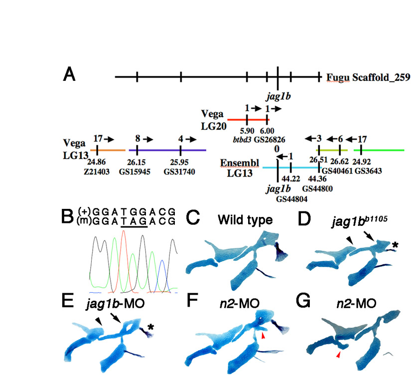
Fig. S1 Identification of the jag1bb1105 mutation. (A) Using synteny between Fugu and zebrafish Vega and Ensembl contigs, the b1105 mutation was mapped to a small region of LG13 containing jag1b. Recombinants per 2000 meioses are shown above each contig, and positions in Mb and marker names below. (B) An electrophoretogram showing sequence surrounding the G-to-A transition that creates a premature stop codon (underlined) in jag1bb1105 mutants (m). (C-G) Unilateral flat-mount dissections of 6 dpf facial skeletons stained for cartilage (blue) and bone (red). jag1b-MO larvae show similar skeletal defects to jag1bb1105 mutants, including truncation of Pq (arrowheads), shape changes in Hm (arrows) and transformed Op bone (asterisks). In notch2-MO larvae, ectopic cartilage processes (red arrowheads) are seen at low frequency near the DV interfaces of the hyoid (F) and mandibular (G) skeletons.