
Fig. 6
- ID
- ZDB-IMAGE-100504-6
- Genes
- Antibodies
- Publication
- Tay et al., 2010 - A vertebrate-specific Chp-PAK-PIX pathway maintains E-cadherin at adherens junctions during zebrafish epiboly
- All Figures
- Figures for Tay et al., 2010
|
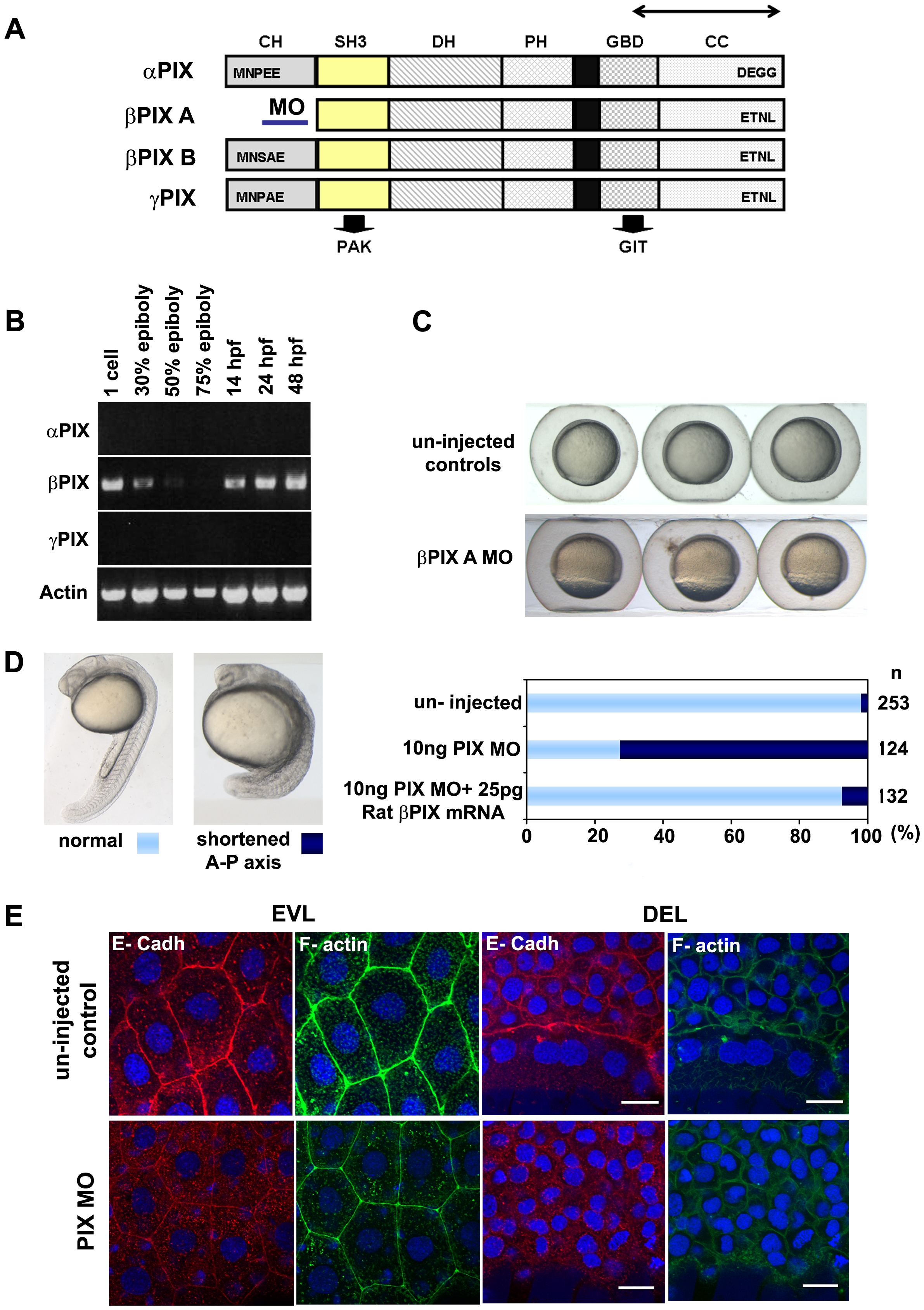
Fig. 6 PIX is required to localize E-cadh at the AJs.
(A) Schematic of zebrafish PIX isoforms designated αPIX, βPIX-A, βPIX-B [7] and the newly described γPIX. Arrows indicate the positions of the oligonucleotide primers used for RT-PCR. The position of the PIX-MO at the 5′ UTR of βPIX-A is indicated: this transcript encodes the smaller PIX isoform which is equivalent to the ubiquitous mammalian βPIX. SH3 domain in yellow and GBD binds to PAK and GIT respectively. (B) Transcript profile showing RT-PCR products for PIX at the developmental stages indicated. Primers cover essentially the same region of the PIX ORFs and therefore do not discriminate between the alternate spliced forms at the 5′ terminus. (C) PIX morphant embryos exhibit epibolic delay compared to un-injected controls at 8 hpf. (D) The typical phenotype of embryos depleted of βPIX-A at 24 hpf, exhibit shortened AP axes suggesting gastrulation defects. Phenotypic analysis showing significant rescue at 24 hpf after co-injection with 25 pg of rat βPIX mRNA. (E) Reduced cell junctional E-cadh signals in the EVL and DEL after PIX knock-down. The level of cortical F-actin (phalloidin) is similar to controls but the junctional network is more irregular. Intracellular E-cadh puncta suggest PIX functions downstream of Chp to maintain E-cadh at cell adhesions. Scale bars = 20 μm.