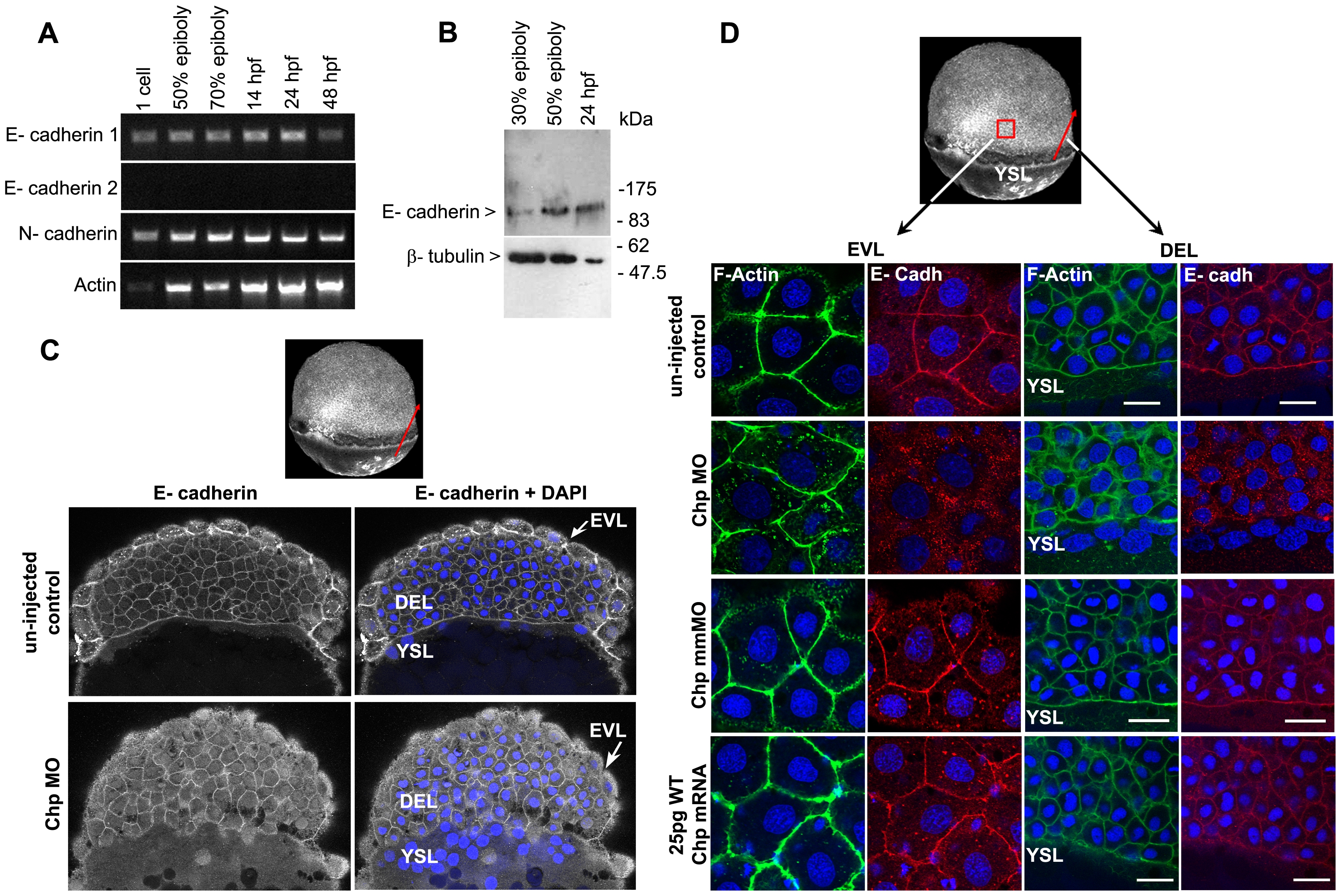
Fig. 4 E-cadh is not maintained at the AJs in the absence of Chp.
(A) RT-PCR products of zebrafish E-cadh1, E-cadh2 and N-cadherin (N-cadh) at the stages shown. E-cadh1 transcript was present throughout all stages however we did not detect expression of E-cadh2 (although it is represented 3 times in the EST database). N-cadh transcripts accumulate more significantly at 50% epiboly. Primers encoding the cytoplasmic domain of E-cadh1 NM_131820, E-cadh2 XM_690906 and N-cadh AF 418565 were used. (B) Immunoblot analysis of endogenous E-cadh at the stages indicated. The E-cadh antibody (BD Biosciences) detects a single E-cadh band (approx. 120kDa). (C) Low resolution image of phalloidin stained embryo of 60% epiboly, marked with red arrow representing the ‘y’ component in xy cross-section of the embryo (EVL→ DEL→ YSL) where the confocal image is taken. A middle sagittal plane of the embryo at 60% epiboly is derived from the Z stack. E-cadh was no longer maintained at the AJs in the EVL and was found predominately in cytoplasm. (D) Schematic diagram (3D view) of 60% epiboly. Red box indicates the lateral area of EVL and red arrow indicates the ‘y vector’ of the xy confocal slice. Confocal images (40x mag.) showing E-cadh co-localized with F-actin at the AJs between adjacent cells in the EVL and DEL. These observations were similar with the controls that were injected with 25 pg WT Chp mRNA alone and Chp mmMO. The images represent a stack of 10 images (each 0.5 μm); E-cadh staining was rarely detected at AJs in Chp morphant but intracellular signal was not diminished, suggesting E-cadh is mis-localized without Chp signaling. Identical loss of E-cadh was observed (data not shown) with a Mab that recognized the extracellular domain of E-cadh (ECM Biosciences; CM1681). Scale bars represent 20 μm.

