
|
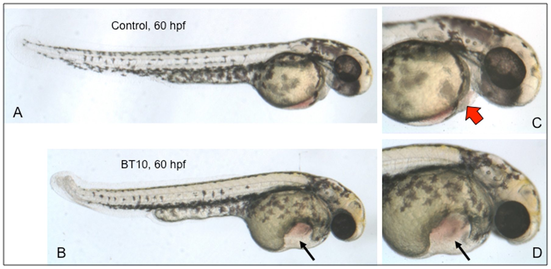
Fig. 4 BT10 causes specific cardiovascular defects when added at 24 hpf.
Shown are representative embryos (from 3 experiments, for each sample n = 20) that were cultured between 24–60 hpf in the presence of DMSO alone (A, control) or BT10 (B). Note the defined morphological structure of the ventral yolk sac (arrow). A normal looping heart tube fails to develop in these embryos. Panels C and D show higher magnification views of representative control (C) and BT10-treated (D) embryos, respectively. The red block arrow indicates blood flowing through the normal control heart, which is not evident in the BT10-treated embryos. Views are lateral, anterior to the right, dorsal at the top.