
Fig. 4
- ID
- ZDB-IMAGE-100429-71
- Genes
- Publication
- Pretorius et al., 2010 - Identification and functional analysis of the vision-specific BBS3 (ARL6) long isoform
- All Figures
- Figures for Pretorius et al., 2010
|
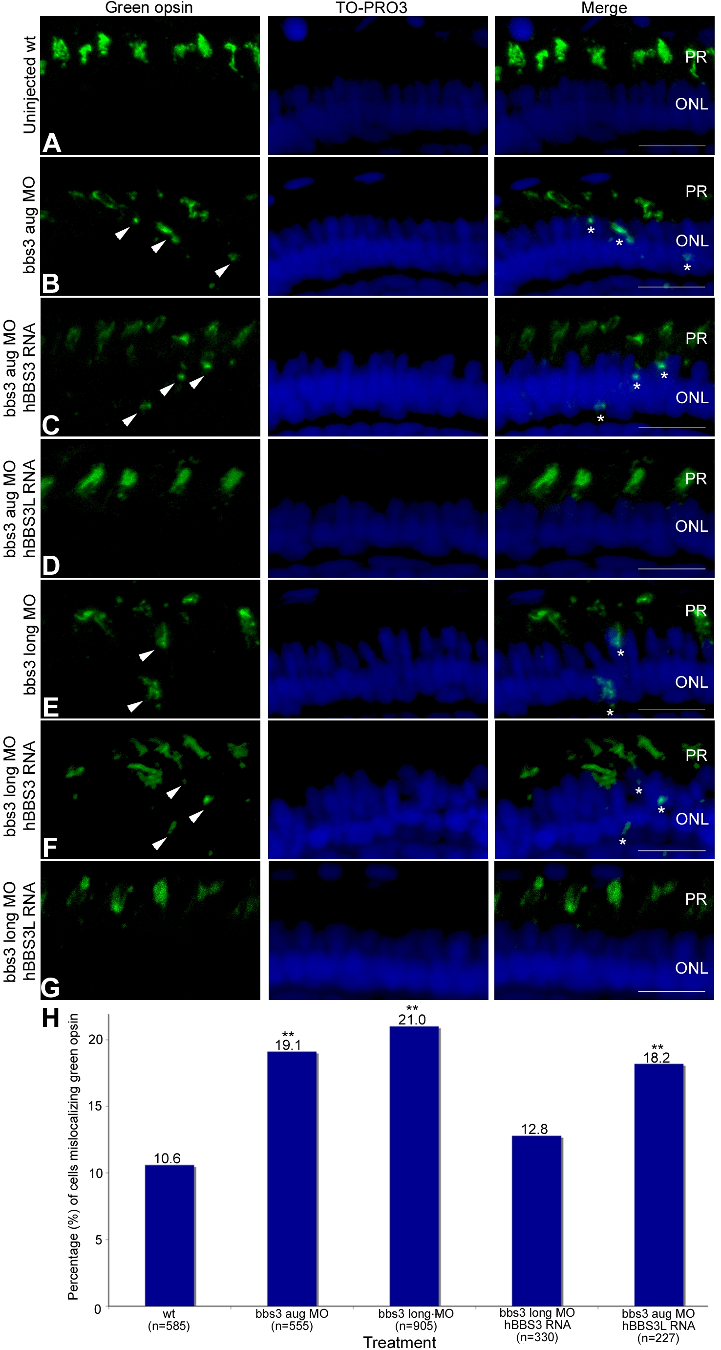
Fig. 4 Green opsin mislocalization and rescue in 5-day-old bbs3 morphant zebrafish.
Immunofluorescence of green cone opsin (green) on transverse cryosections of 5-day-old embryos (A) uninjected wild-type, (B) bbs3 aug MO, (C) bbs3 aug MO and hBBS3 RNA, (D) bbs3 aug MO and hBBS3L RNA, (E) bbs3 long MO, (F) bbs3 long MO and hBBS3 RNA, and (G) bbs3 long MO and hBBS3L RNA. To-Pro3 was used to counterstain the nuclei (blue). In bbs3 aug (B) and bbs3 long morphants (E), green opsin was not restricted to the outer segment of the photoreceptors and was detected in the cell bodies of the outer nuclear layer (arrowheads and asterisks). Expression of hBBS3L RNA improved green opsin localization in both bbs3 aug (D) and bbs3L (G) morphants. Of note, hBBS3 RNA failed to rescue green opsin localization in bbs3 aug (C) and bbs3 long (F) morphant embryos (arrowheads and asterisks). OS, outer segment; ONL, outer nuclear layer. Scale Bar 10 µm. (H) The percentage of mislocalizing green opsin cells. The sample size (n) is noted on the x-axis and represents the total number of green opsin positive cells counted. **Fisher′s exact test, p<0.01.