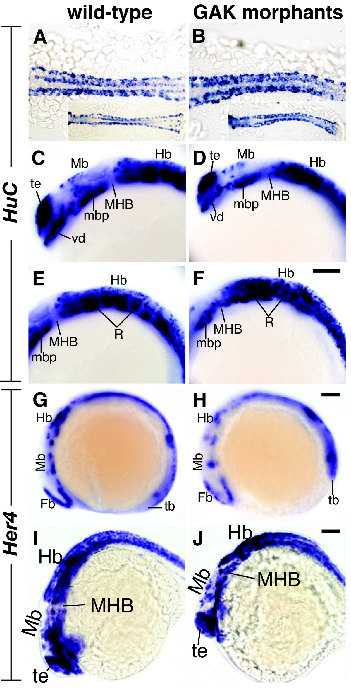
Fig. 10 Expression patterns of HuC and Her4 in wild-type and GAK morphant embryos. (A, B) Close-up dorsal views of HuC expression in (A) wild-type and (B) GAK morphant embryos at the 8-somite stage. Micrographs of a lower magnification are shown in the insets. At this stage, more cells appeared to express HuC in GAK morphant embryos, suggesting the presence of more neural progenitor cells. (C-F) Lateral views of HuC expression in the brain regions of (C, E) wild-type and (D, F) GAK morphant embryos at 24 to 28 hpf. A comparison of (C) and (D) shows a reduction in HuC-positive cells in the forebrain and midbrain in GAK morphants. Similarly, a comparison of (E) and (F) shows the disorganization and reduction of HuC-positive cells in the hindbrain in GAK morphants. (G-J) Lateral views of Her4 expression patterns in (G, I) wild-type and (H, J) GAK morphants at the (G, H) 8-somite stage or at (I, J) 24 to 28 hpf. At the 8-somite stage, Her4 expression in GAK morphant embryos was significantly reduced, as compared to the wild type. In contrast, the expression of Her4 in the brain of wild-type and GAK morphant embryos at 24 to 28 hpf was comparable. In all the panels, anterior is to the left, and in all the lateral views, dorsal is up. Fb, forebrain; Hb, hindbrain; MHB, mid-hindbrain boundary; Mb, midbrain; mbp, midbrain basal plate; R, rhombomeres; tb, tailbud; te, telencephalon; vd, ventral diencephalon. Scale Bar, 100 μm.
Image
Figure Caption
Figure Data
Acknowledgments
This image is the copyrighted work of the attributed author or publisher, and
ZFIN has permission only to display this image to its users.
Additional permissions should be obtained from the applicable author or publisher of the image.
Full text @ BMC Dev. Biol.

