
|
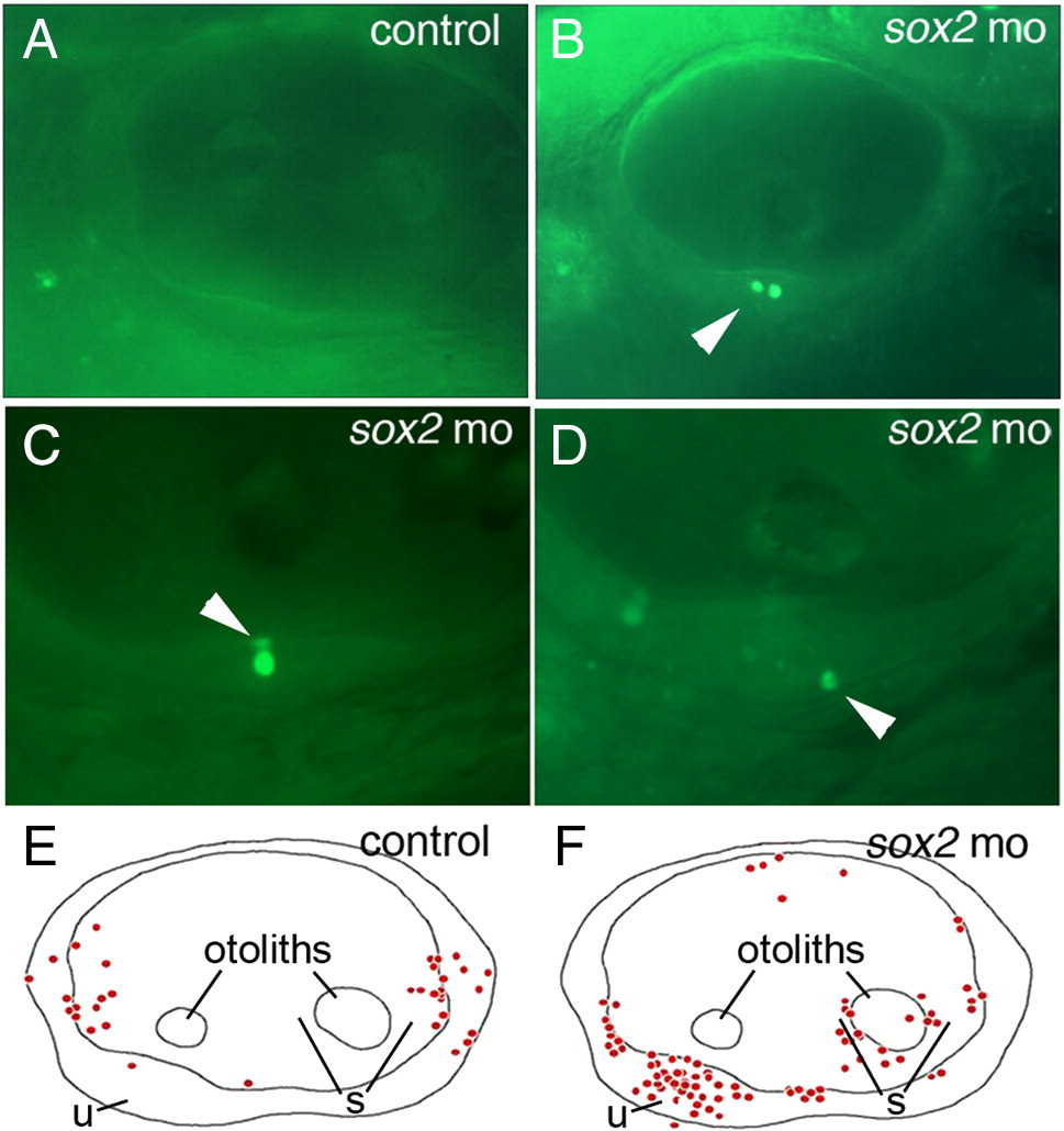
Fig. 2 Loss of Sox2 results in macular death. (A–D) AO-labeling of dying cells in a control embryo (A) and sox2 morphants (B–D). Morphants often contained multiple dying cells within sensory epithelia (B), and were observed in apical (C) or basal (D) regions of the maculae (arrowheads). (E and F) Schematic maps depicting the distribution of all AO-positive cells seen in otic vesicles of 33 control embryos (E) or 33 sox2 morphants (F) at 48 hpf. Positions of the utricular macula (u), saccular macula (s) and otoliths are indicated. No AO-positive cells were detected in the lateral wall of the otic vesicle. All images show lateral views with anterior to the left and dorsal to the top.
Reprinted from Developmental Biology, 338(2), Millimaki, B.B., Sweet, E.M., and Riley, B.B., Sox2 is required for maintenance and regeneration, but not initial development, of hair cells in the zebrafish inner ear, 262-269, Copyright (2010) with permission from Elsevier. Full text @ Dev. Biol.