
Fig. S5
- ID
- ZDB-IMAGE-100225-10
- Publication
- Sun et al., 2010 - Zili Inhibits TGF-beta Signaling by Interacting with Smad4
- All Figures
- Figures for Sun et al., 2010
|
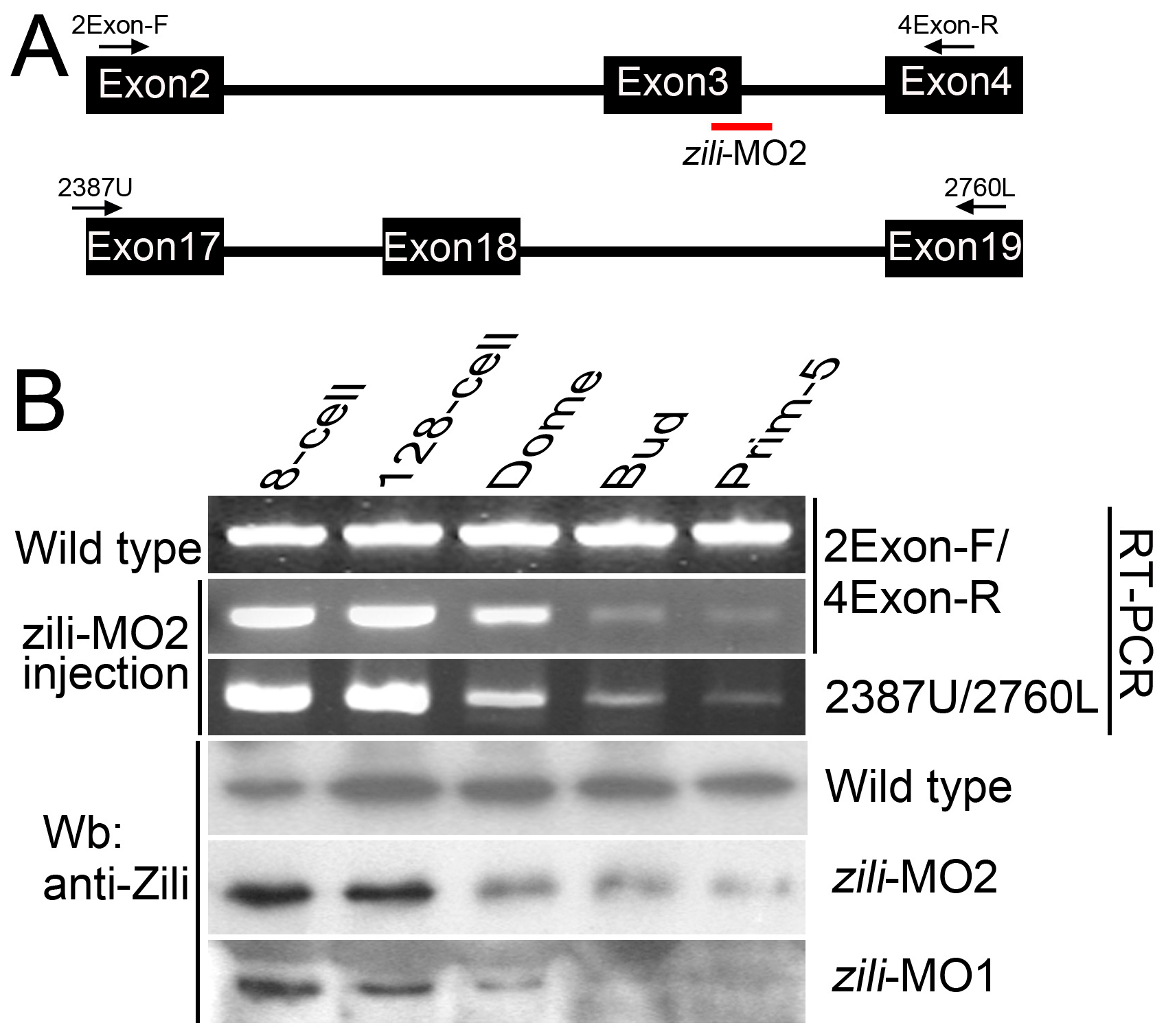
Fig. S5 Effectiveness of the splice-inhibiting morpholino (zili-MO2). (A) Parts of genomic structure of zili gene. Exons and primers are indicated. Splice site targeted by zili-MO2 is shown. (B) RT-PCR and Western blotting assays to detect the effectiveness of zili-MO2 and zili-MO1. Detected by primer pair 2Exon-F/4Exon-R, injection of zili-MO2 produces diminution of the wild type transcript band beginning at the Dome stage (4.3 hpf), a point after the initiation of zygotic transcription (Line 2), suggesting that zili-MO2 specifically target zygotic, and not maternal, transcripts (1). Furthermore, the products amplified by 2Exon-F/4Exon-R were identified by sequencing. Another primer pair 2387U/2760L, targeting to Exon 17 and 19 outside of the region expected to be modified by zili-MO2, also detects the diminution (Line 3), suggesting that the entire zili transcript decreases due to zili-MO2. Consistent with the results of RT-PCR, diminution of Zili protein induced by zili-MO2 and 1 can also be detected by western blotting (Line 5 and 6). Zili-MO2, locating at the exon3-intron3 boundary, was designed by Gene Tools, LLC and would generate an out-of-frame mutation resulting in a loss-of-function phenotype. Injection of zili-MO2 can produce pre-mRNA sequence with a premature termination codon brought in-frame by the exon3 excision and the pre-mRNA will undergo nonsense-mediated decay (2). So the splice variants can not be detected.