
|
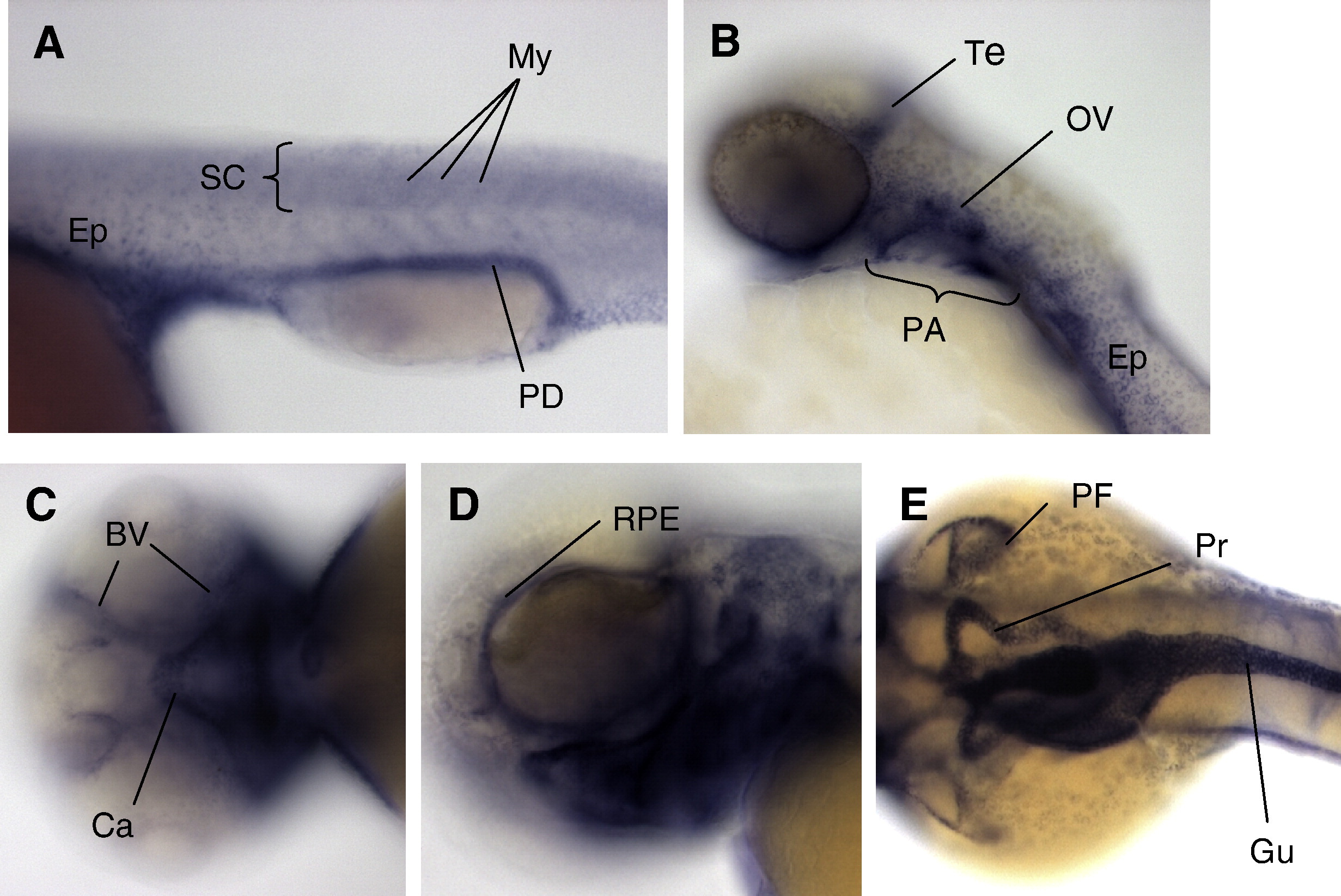
Fig. 1 Zebrafish col18a1 displays a complex and dynamic development expression pattern. Expression of col18a1 was detected by whole embryo in situ hybridization at 24 hpf (A); 2 dpf (B); and 3 dpf (C–E). Dynamic expression was seen in neutral structures including spinal cord (A) and tectum (B). Expression was also seen in pronephric duet and pronephros (A, E); myospetum (A); epidermis (A, B); otic vesicle (B); pharyngeal arches (B); craniofacial cartilages and blood vessels (C); retinal pigment epithelium (D); pectoral fins (E); and gut endothelium (E). Abbreviations used in all figures: BV (blood vessels), Ca (cartilage), FB (forebrain), Gu (gut endothelium), My (myoseptum), OV (otic vesicle), PA (pharyngeal arches), PD (pronephric duct), PF (pectoral fin), Pr (pronephros), Re (retina), RPE (retinal pigment epithelium), SC (spinal cord), Te (tectum).
Reprinted from Developmental Biology, 337(2), Kague, E., Bessling, S.L., Lee, J., Hu, G., Passos-Bueno, M.R., and Fisher, S., Functionally conserved cis-regulatory elements of COL18A1 identified through zebrafish transgenesis, 496-505, Copyright (2010) with permission from Elsevier. Full text @ Dev. Biol.