
Fig. 4
- ID
- ZDB-IMAGE-100128-19
- Genes
- Publication
- Mitchell et al., 2010 - Effect of Vascular Cadherin Knockdown on Zebrafish Vasculature during Development
- All Figures
- Figures for Mitchell et al., 2010
|
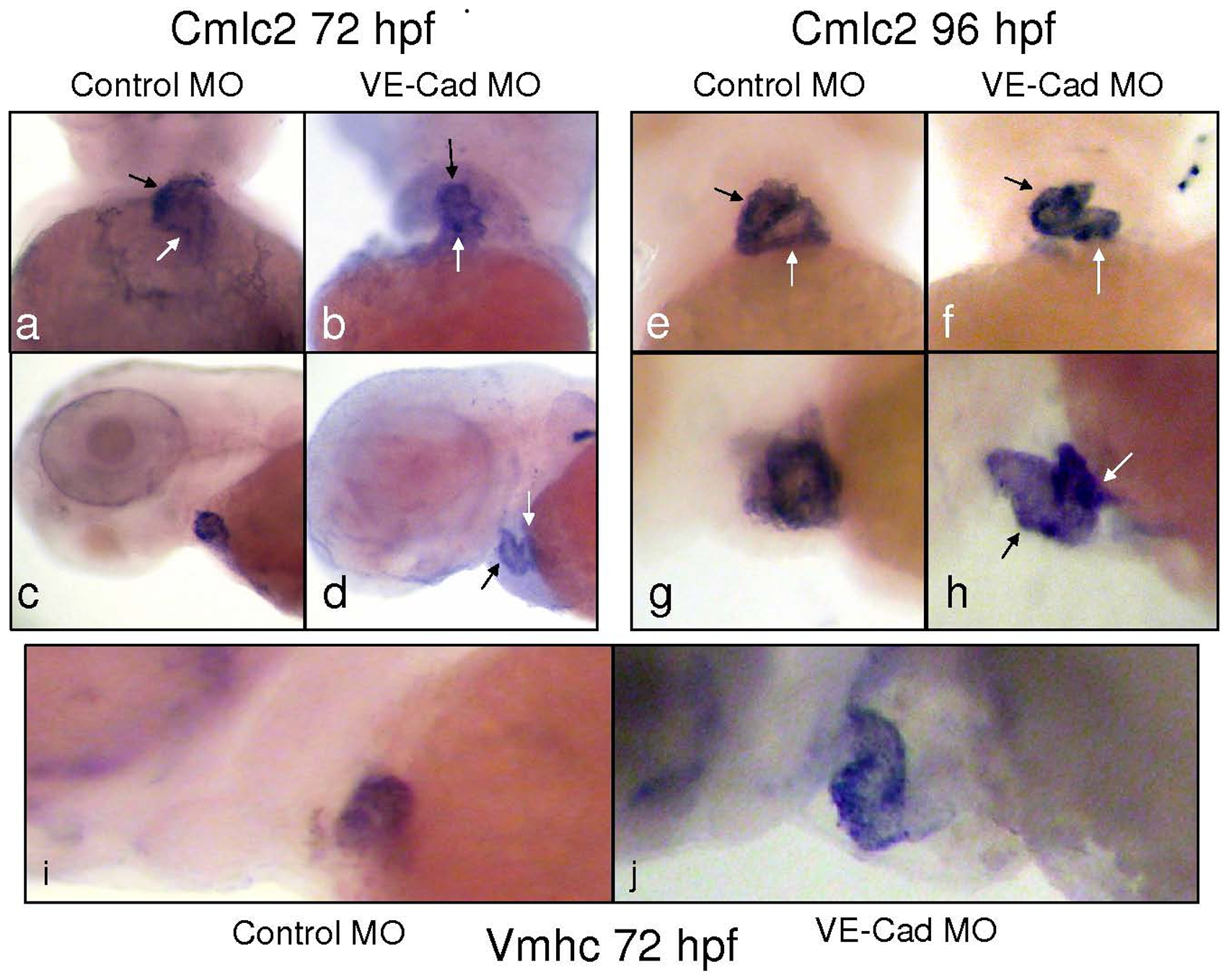
Fig. 4 Knockdown of VE-cadherin causes defective cardiac looping and chamber thinning.
In situ hybridization of Control MO and VE-cad MO injected embryos with the pan-cardiac myosin marker cmlc2 (a–h) and the ventricular-specific vmhc (i–j). Ventral and lateral images of control embryos stained for cmlc2 (a,c) show proper atrial (white arrow) and ventricular (black arrows) looping. Images of VE-Cad knockdown embryos show a linear arrangement of the cardiac chambers (b,d). By 96 hours, cmlc2 staining shows the normal side-by-side arrangement of chambers in control embryos (e,g), while the heart remains thinned and tubular in VE-cad MO injected embryos (f,h). Staining for vmhc at 72 hours shows a compact ventricular chamber in controls (i), while the ventricles of knockdown embryos are elongated and thinner-appearing (j).