
Fig. 4
- ID
- ZDB-IMAGE-100121-6
- Publication
- O'Brien et al., 2009 - Developmentally Regulated Impediments to Skin Reinnervation by Injured Peripheral Sensory Axon Terminals
- All Figures
- Figures for O'Brien et al., 2009
|
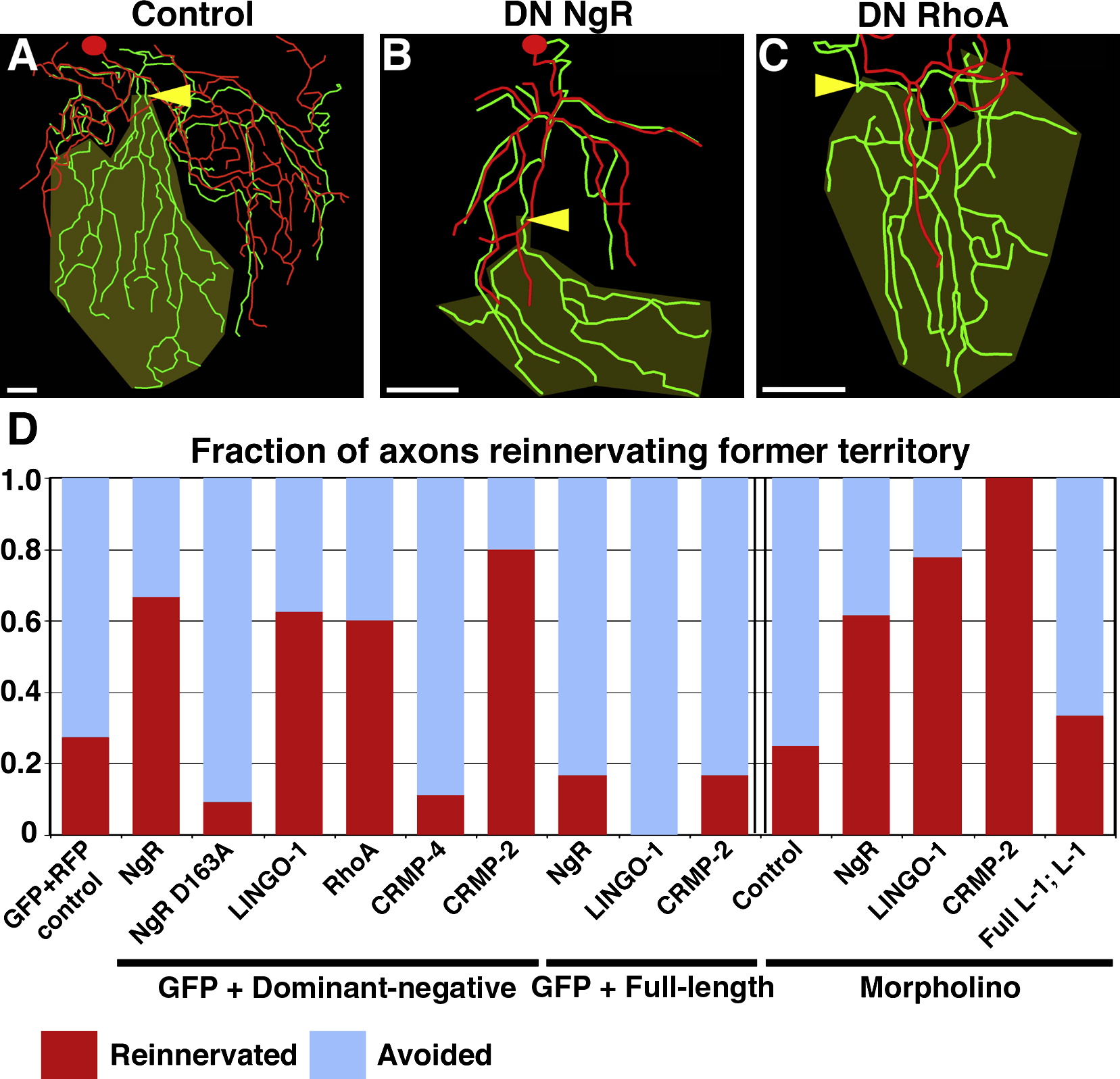
Fig. 4 Inhibition of Skin Reinnervation by Injured Axons Is Mediated by the NgR/RhoA Pathway
(A–C) Tracing overlays as in Figure 2. Arrowhead is site of axotomy, olive marks denervated territory, and scale bars represent 50 μm. Axotomy at 78 hpf of a trigeminal neuron expressing GFP and RFP (A), dominant-negative (DN) NgR (B), or DN RhoA (C) is shown.
(D) Quantification of fraction of axons that entered the denervated territory. Data to the left of the double bar are from axons expressing GFP and dominant-negative or full-length versions of the genes indicated. Data to the right of the double bar are from Tg(sensory:GFP) embryos injected with indicated morpholinos. Red indicates fraction of axons that grew into denervated territory; blue indicates axons that avoided denervated territory (see Supplemental Experimental Procedures). See Table S2 and Movies S11–S13.