
Fig. 2
- ID
- ZDB-IMAGE-091215-63
- Genes
- Publication
- Lee et al., 2009 - Notch Signaling Functions as a Cell-Fate Switch between the Endothelial and Hematopoietic Lineages
- All Figures
- Figures for Lee et al., 2009
|
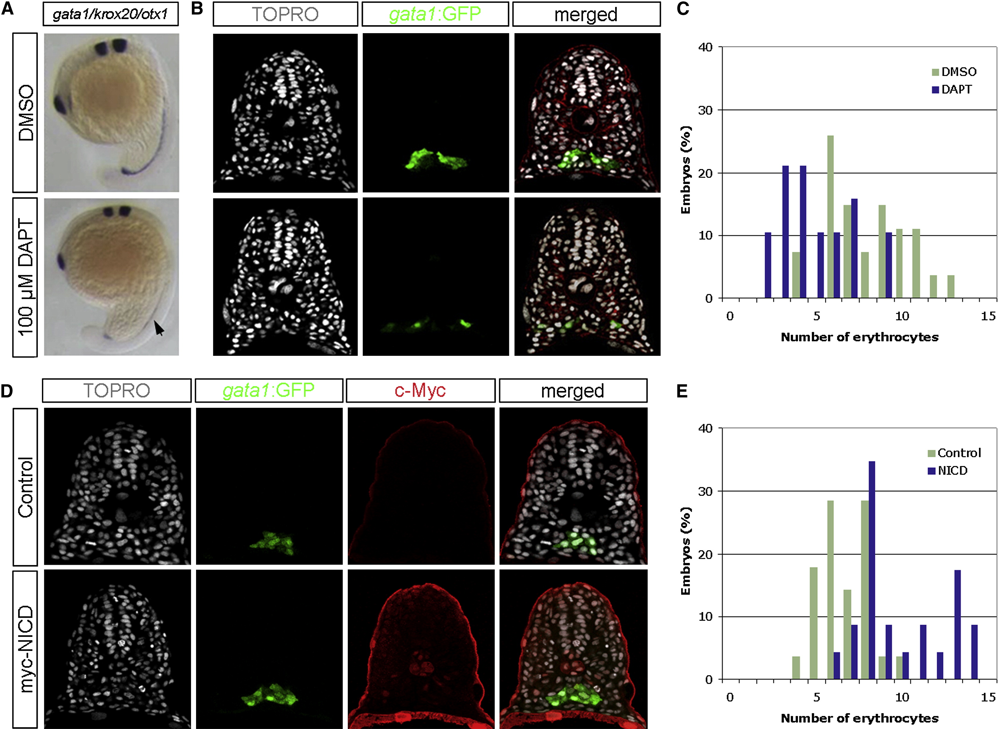
Fig. 2 Notch Signaling Positively Regulates the Number of Hematopoietic Cells during Zebrafish Development
(A) Whole-mount RNA in situ hybridization showing the expression of gata1 in 18 hpf DMSO-treated or DAPT-treated embryos. Two additional markers, krox20 and otx1, which label the hindbrain and neuroectoderm, respectively, were used as positive controls. Notice the reduced expression of gata1 in DAPT-treated embryos (black arrow).
(B) Transverse sections of 18 hpf DMSO- or DAPT-treated embryos visualized for TOPRO (white), gata1:GFP (green), and phalloidin (red).
(C) Quantification of the hematopoietic nuclei per focal plane in DMSO-treated (n = 27) or DAPT-treated (n = 19) embryos.
(D) Transverse sections of 18 hpf phenotypic wild-type siblings and Tg(hsp70l:Gal4)kca4;Tg(UAS:myc-NICD)kca3 embryos visualized for TOPRO (white), gata1:GFP (green), and c-Myc as a surrogate measure for NICD expression (red).
(E) Quantification of the hematopoietic nuclei per focal plane in phenotypic wild-type siblings (n = 28) and NICD-overexpressing embryos (n = 23). DAPT-treated embryos contained 4.89 (s = 2.15) erythrocyte nuclei, in comparison to 8.07 (s = 2.38) nuclei in DMSO-treated embryos (ANOVA, p < 10-4). Conversely, embryos overexpressing the NICD contained 9.89 (s = 2.53) erythrocyte nuclei, in comparison to 6.72 (s = 1.43) nuclei per section in phenotypic wild-type siblings (ANOVA, p < 10-6), indicating that Notch signaling positively modulates the number of hematopoietic cells.