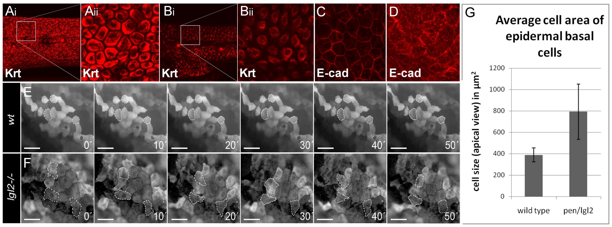
Fig. 2 Epidermal cells undergo EMT in pen/lgl2 mutant larvae.
Basal epidermal cells of wild type and pen/lgl2 mutant 5dpf stained using pan 1–8 Cytokeratin antibody (Ai - Bii) and E-cad antibody (C, D). Time lapse analysis of tg(ΔNp63::Gal4,UAS::GFP) labelled cells in wild-type (E) and pen/lgl2 mutant larvae (F) at 5dpf. Analysis of cell area in wild-type and pen/lgl2 mutant larvae (G). In contrast to wild-type larvae (Ai, Aii), in pen/lgl2 mutant larvae the cells appear spindle shaped with keratin accumulation around the nucleus (Bi, Bii). Furthermore, in wild-type basal epidermal cells (C), E-cad localizes to the cell membrane; cells exhibit perfect polygonal shapes. In pen/lgl2 mutant larvae (D) membrane localization of E-cad is strongly reduced with concomitant increase in the cytoplasmic fraction. Time-lapse analysis reveals (E, F) that there is no change in the shapes of wild-type epidermal cells (E). However, in pen/lgl2 mutants (F) the shape of epidermal cells dramatically changes over time, indicating their metastable cell fate. Cells develop lamellipodia like structures, a classic trait exhibited by mesenchymal cell types (see Videos S1, S2, S3, S4 in addition). As epidermal basal cells flatten and develop lamellipodia like cell protrusions in pen/lgl2, the cell area in apical view is increased in these larvae (G). However, average area of epidermal basal cells in these mutants is highly variable compared to wild types.

