
Fig. S3
- ID
- ZDB-IMAGE-091121-9
- Publication
- Pittman et al., 2008 - Pathfinding in a large vertebrate axon tract: isotypic interactions guide retinotectal axons at multiple choice points
- All Figures
- Figures for Pittman et al., 2008
|
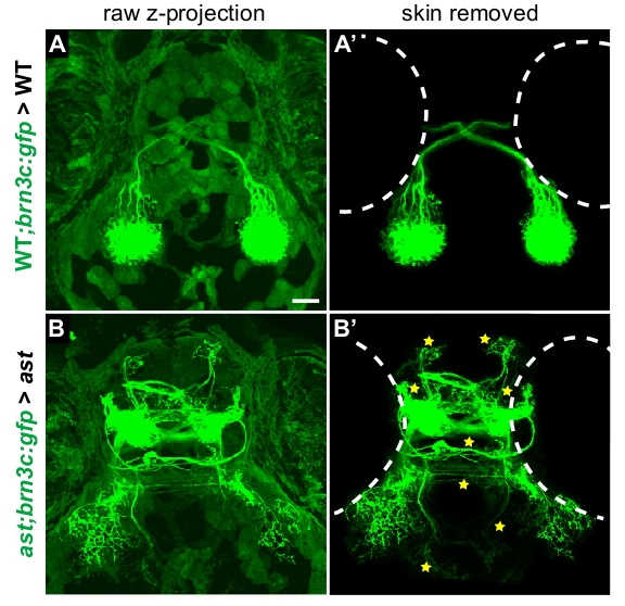
Fig. S3 Background removal procedure used for transplant images. Dorsal views of projections made by donor axons in 5 dpf host larvae, control transplants. (A,B) Maximum-intensity z-projections of unedited confocal stack in wild type>wild type (A) and ast>ast (B) transplants. Autofluorescence in skin, superficial to axons, partially obscures the retinal projection. (A′,B′) Confocal projections shown after removing autofluorescence slice-by-slice by manual editing in ImageJ and Adobe Photoshop. Retinal axons are unaffected, but can be seen more clearly in the absence of skin fluorescence. Stars indicate errors (always counted in unedited confocal stacks); dotted outlines show eye positions. All transplant images in Figs 5 and 6 were processed in this manner.