
Fig. S2
- ID
- ZDB-IMAGE-091016-21
- Publication
- Emond et al., 2009 - Protocadherin-19 is essential for early steps in brain morphogenesis
- All Figures
- Figures for Emond et al., 2009
|
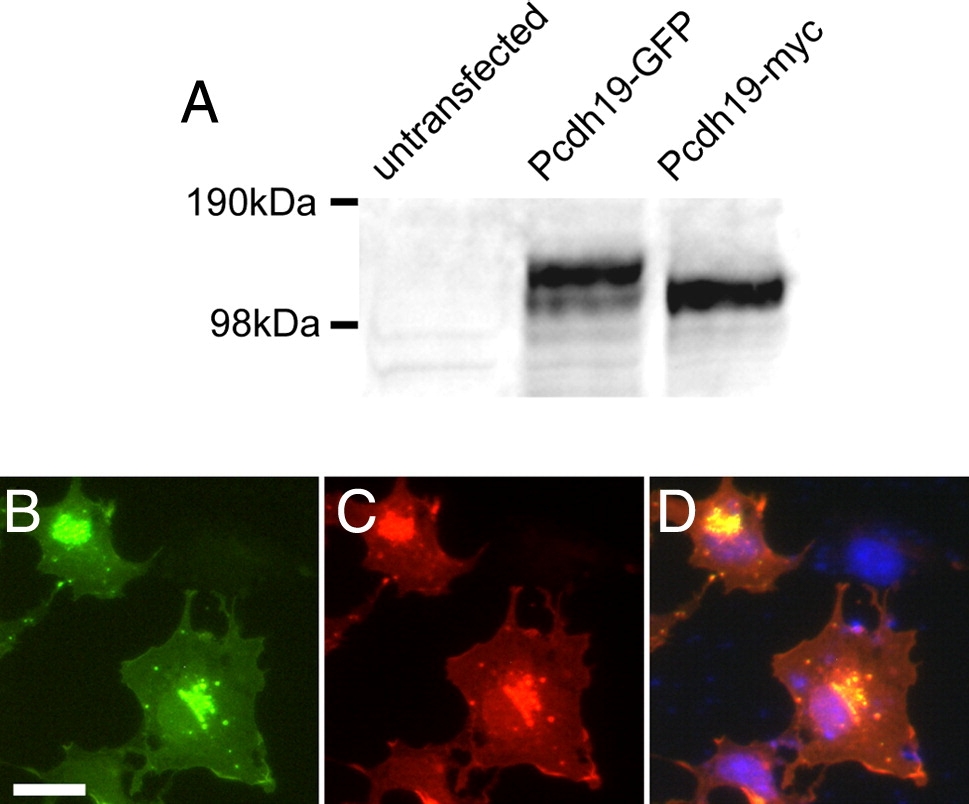
Fig. S2 Specificity of anti-Pcdh19 antibody. A, Shown is a Western blot of cell extracts from COS cells that were untransfected or transfected with Pcdh19-GFP or Pcdh19-myc. In untransfected cells, no band is present, while in transfected cells, our antibody detects strong bands corresponding to the sizes of Pcdh19-GFP and Pcdh19-myc, respectively. B–D, Immunocytochemistry of COS cells transfected with zebrafish Pcdh19-GFP. B, Pcdh19-GFP is present in a perinuclear compartment and diffusely along the plasma membrane, as well as in discrete, punctate structures. C, Antibody labeling shows an identical distribution to the Pcdh19-GFP. D, An overlay shows excellent co-localization of Pcdh19-GFP (green) and anti-Pcdh19 (red), while there is no immunolabeling of untransfected cells in the same field (DAPI, blue).
Reprinted from Developmental Biology, 334(1), Emond, M.R., Biswas, S., and Jontes, J.D., Protocadherin-19 is essential for early steps in brain morphogenesis, 72-83, Copyright (2009) with permission from Elsevier. Full text @ Dev. Biol.