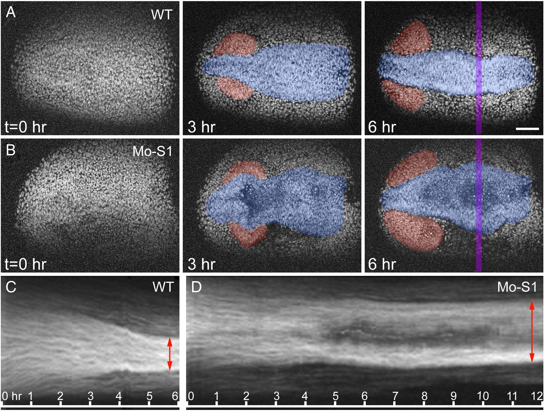
Fig. 10 In vivo timelapse imaging reveals an arrest of convergence movements during neurulation in pcdh19 morphants. (A) Images extracted from a 6 hour timelapse sequence of transgenic embryos expressing a Histone H2A-GFP fusion protein. Each frame is a maximum intensity projection of 20 optical sections (spaced at 5 μm). Shown are three timepoints, at 3 hour intervals, revealing a net migration of cells toward the midline. By 3 h after the initiation of the timelapse, eye primordia have begun to form (red transparency), and the embryo has begun to narrow mediolaterally (blue transparency). Anterior is to the left; scale bar = 50 μm. (B) Images extracted from a 12 hour timelapse of transgenic embryos that have been injected with Mo-S1. In contrast to wild type embryos, cell movement toward the midline is arrested. (C) A kymograph from the 6 hour timelapse image sequence in A, showing the cell convergence. The increase in fluorescence intensity reflects an increase in cell density. The kymograph shows the intensity values measured along a line (11 pixel width, shown as a transparent purple bar in A) at each timepoint. (D) A kymograph from the timelapse image sequence in B, showing a failure of cell convergence. This data was assembled from a 12 hour timelapse sequence. Movement of cells toward the midline is disrupted in pcdh19 morphants, resulting in a lateral accumulation of cells. Compare red arrows in C and D.
Reprinted from Developmental Biology, 334(1), Emond, M.R., Biswas, S., and Jontes, J.D., Protocadherin-19 is essential for early steps in brain morphogenesis, 72-83, Copyright (2009) with permission from Elsevier. Full text @ Dev. Biol.

