
Fig. 5
- ID
- ZDB-IMAGE-090817-5
- Genes
- Antibodies
- Publication
- McMahon et al., 2009 - Lmx1b is essential for survival of periocular mesenchymal cells and influences Fgf-mediated retinal patterning in zebrafish
- All Figures
- Figures for McMahon et al., 2009
|
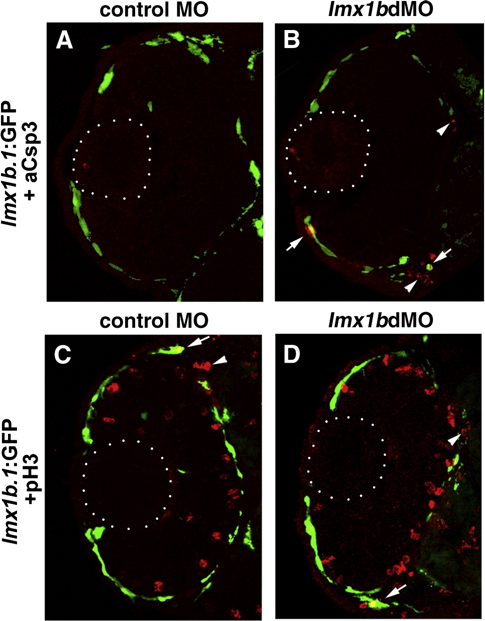
Fig. 5 Apoptosis and proliferation in lmx1b.1:GFP-positive cells following loss of Lmx1b function. (A) Immunoreactivity for activated Caspase-3 (red) and fluorescence for lmx1b.1:GFPmw11 (green) in control MO or (B) lmx1bdMO ocular cryosections at 36 hpf. Arrows indicate cells positive for both activated Caspase-3 and lmx1b.1:GFP, while arrows indicate GFP-negative apoptotic cells. (C) Immunoreactivity for phosphoHistone (red) and fluorescence for lmx1b.1:GFPmw11 (green) in control MO or (D) lmx1bdMO ocular cryosections at 36 hpf. Arrows indicate cells positive for both activated phosphoHistone-3 and lmx1b.1:GFP, while arrows indicate GFP-negative mitotic periocular cells. Dotted lines indicate position of the lens. Images shown are from representative sections (n = 24 total embryos for each condition from 2 experiments).
Reprinted from Developmental Biology, 332(2), McMahon, C., Gestri, G., Wilson, S.W., and Link, B.A., Lmx1b is essential for survival of periocular mesenchymal cells and influences Fgf-mediated retinal patterning in zebrafish, 287-298, Copyright (2009) with permission from Elsevier. Full text @ Dev. Biol.