
Fig. 2
- ID
- ZDB-IMAGE-090814-1
- Genes
- Publication
- Feijoo et al., 2009 - Cystein-serine-rich nuclear protein 1, Axud1/Csrnp1, is essential for cephalic neural progenitor proliferation and survival in zebrafish
- All Figures
- Figures for Feijoo et al., 2009
|
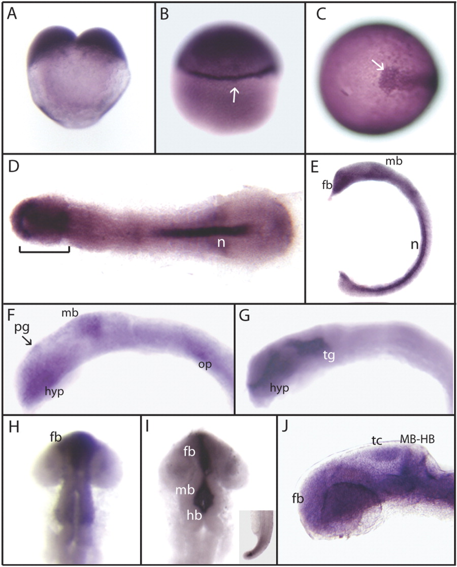
Fig. 2 Expression of zebrafish axud1 gene. Detection of axud1 mRNA was carried out by whole-mount in situ hybridization using different stage embryos from two cells to 24 hpf. Images in A, B, E, F, G, and J are lateral views, C is a dorso-anterior view, and D, H, and I are dorsal views. A: Two-cell-stage embryo. B: Thirty percent epiboly. axud1 is detected ubiquitously with higher expression at the marginal zone (arrow) and animal pole. C: A 10-hpf embryo. Expression is detected in the prechordal plate and chordamesoderm, the anterior limit is indicated (arrow). D: A 10-hpf flat-mount embryo. axud1 expression localises in the head and polster regions (bracket) and notochord (n). E: A 12-hpf embryo. Expression is observed in the anterior forebrain, midbrain, and notochord. F: A 16-hpf embryo. axud1 mRNA is found in the anterior hypothalamus, pineal gland (arrow), dorsal midbrain region, and scattered cells in the otic placode. At 20 hpf (G, H) and 22 hpf (I), the expression becomes restricted to the forebrain and midbrain brain ventricles and the tip of the tail (inset in I). J: A 24-hpf embryo. axud1 RNA appears in cephalic regions with higher levels at the forebrain, tectum, midbrain-hindbrain boundary, and hindbrain. fb, forebrain; mb, midbrain; hb, hindbrain; n, notochord; tc, tectum; tg, tegmentum; pg, pineal gland; hyp, hypothalamus; MB-HB, midbrain-hindbrain boundary; op, otic placode.