
IMAGE
Fig. 7
- ID
- ZDB-IMAGE-090804-77
- Publication
- Dodd et al., 2009 - The ENTH domain protein Clint1 is required for epidermal homeostasis in zebrafish
- All Figures
- Figures for Dodd et al., 2009
Image
|
Figure Caption
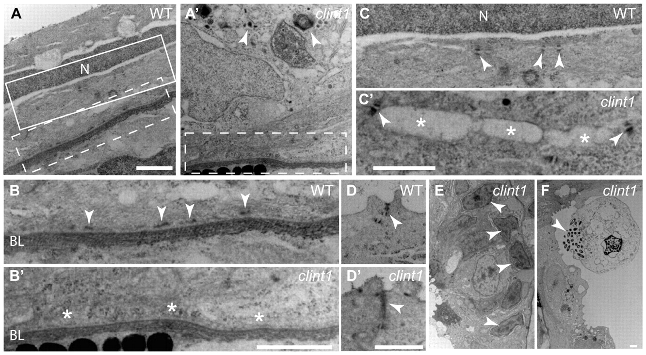
Fig. 7 Impaired hemidesmosome formation in clint1 mutants. Transmission electron micrographs of transverse sections of wild-type (A-D) and clint1 mutant (A′-D′,E,F) zebrafish embryos staged at 4 dpf. The dashed boxed regions in A and A′ are magnified in B and B′, respectively. The solid boxed region in A is magnified in C. Arrowheads label hemidesmosomes (B), desmosomes (C,C′), tight junctions (D,D′), cellular fragmentation (A′,F) and condensed nuclei (E). Asterisks highlight absence of hemidesmosomes (B′) and detachment of epidermal cells (C′). N, nucleus; BL, basal lamina. Scale bars: 1 μm.
Figure Data
Acknowledgments
This image is the copyrighted work of the attributed author or publisher, and
ZFIN has permission only to display this image to its users.
Additional permissions should be obtained from the applicable author or publisher of the image.
Full text @ Development