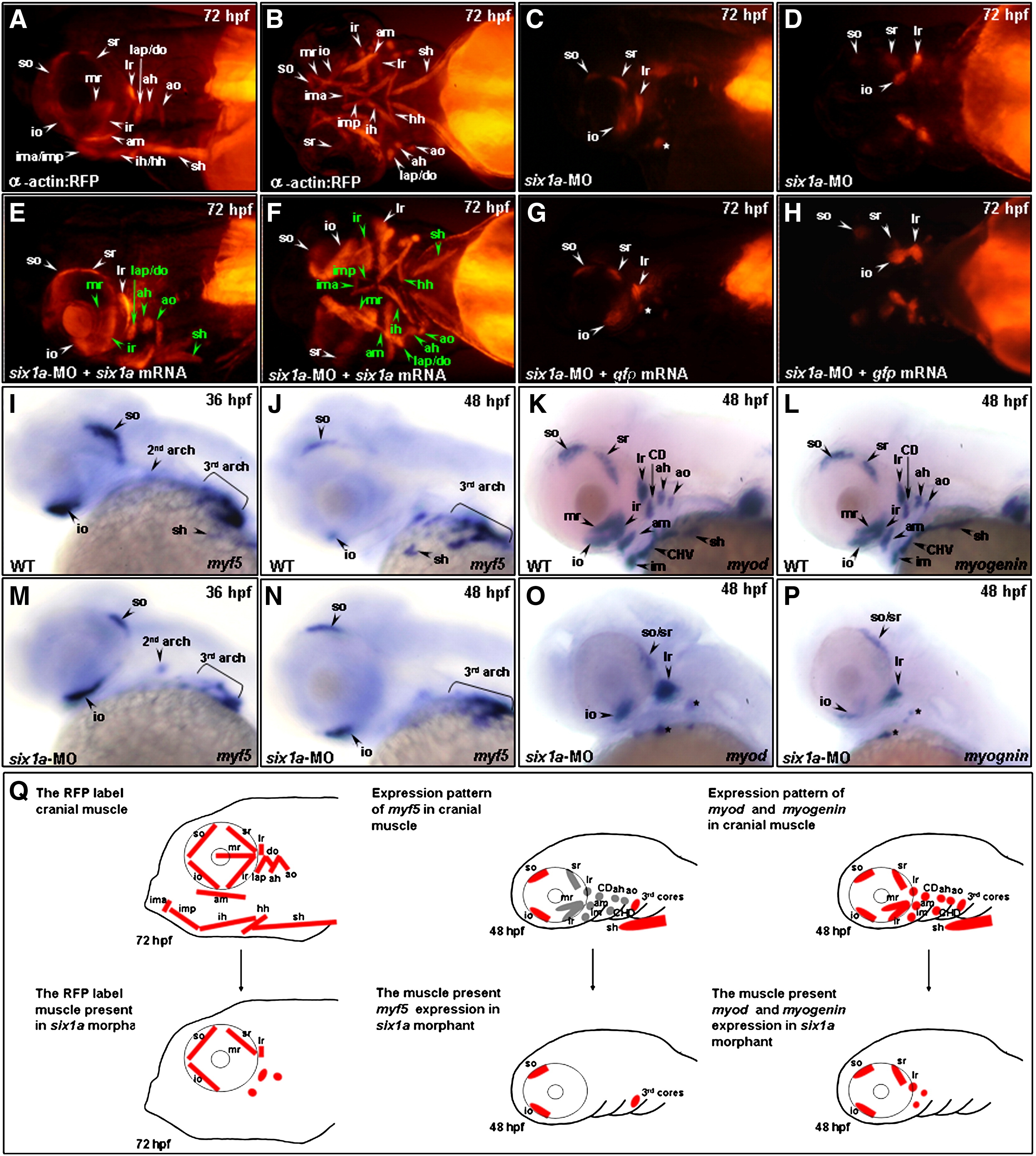
Fig. 2 Six1a is required for the development of mr, ir, sh and all pharyngeal muscles. Embryos derived from the transgenic line Tg(α-actin:RFP) (A–H), all of whose skeletal muscles appear as red fluorescent protein (RFP), were injected with 8 ng of six1a-morpholino oligonucleotide (MO) to specifically inhibit six1a mRNA translation. RFP signal was detected only in the so, io, sr, lr, and remnant dorsal branchial arch muscle (white star) primordia in the six1a-MO-injected embryos (A vs. C and B vs. D). When embryos were injected with 8 ng of six1-MO together with 150 pg of six1a mRNA, results showed that the defective muscle primordia induced by six1a-MO were rescued and appeared as RFP-labeled muscles (E, F; the rescued muscles are marked in green typeface). In contrast, the rescue experiment failed when embryos were injected with 8 ng of six1a-MO with 200 pg of gfp mRNA (G, H), suggesting that the defects of six1a morphants were specific. The expressions of myf5 (I, J, M, N), myod (K, O), and myogenin (L, P) were also observed at the stages indicated. When wild-type embryos were injected with six1a-MO, myf5 was expressed normally in the six1a morphants, both at 36- (I vs. M) and at 48-hpf (J vs. N), except sh. On the other hand, the expressions of myod (K vs. O) and myogenin (L vs. P) were decreased in the extraocular io, so, sr and lr in the six1 morphants at 48 hpf. Weak signals of myod and myogenin were also noticed in the remnant dorsal branchial muscles (black stars) of six1a morphants. The schematic diagram illustrates the cranial muscle defects in six1a morphants and compares the expressions of myf5, myod and myogenin between wild-type (upper row, Q) and six1a morphants (lower row, Q). Lateral view: A, C, E, G and I–P; and ventral view: B, D, F and H. ah, adductor hyoideus; am, adductor mandibulae; ao, adductor operculi; do, dilator operculi; hh, hyohyoideus; ih, interhyoideus; ima, intermandibularis anterior; imp, intermandibularis posterior; io, inferior oblique; ir, inferior rectus; lap, levator arcus palatini; lr, lateral rectus; mr, medial rectus; sh, sternohyoideus; so, superior oblique and sr, superior rectus.
Reprinted from Developmental Biology, 331(2), Lin, C.Y., Chen, W.T., Lee, H.C., Yang, P.H., Yang, H.J., and Tsai, H.J., The transcription factor six1a plays an essential role in the craniofacial myogenesis of zebrafish, 152-166, Copyright (2009) with permission from Elsevier. Full text @ Dev. Biol.

