
Fig. S6
- ID
- ZDB-IMAGE-090710-76
- Publication
- Aanstad et al., 2009 - The Extracellular Domain of Smoothened Regulates Ciliary Localization and Is Required for High-Level Hh Signaling
- All Figures
- Figures for Aanstad et al., 2009
|
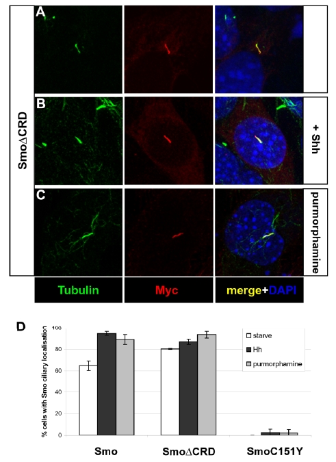
Fig. S6
The ciliary localization of SmoΔCRD is independent of Hh pathway activation.
Expression of acetylated Tubulin (green) and Myc-tagged SmoΔCRD (red) in NIH-3T3 cells (A-C). Nuclei were visualized with DAPI (blue). SmoΔCRD localized to the cilium in the absence of pathway activation (A), as well as in the presence of Shh (B) or purmorphamine (C). (D) Quantification of ciliary localization of wild-type Smo, SmoΔCRD and SmoC151Y in NIH-3T3 cells. For quantification, all transfected cells were assessed, independent of expression level. High level expression of wild-type Smo was observed to be sufficient to promote ciliary localization.