
IMAGE
Fig. S5
- ID
- ZDB-IMAGE-090710-75
- Publication
- Aanstad et al., 2009 - The Extracellular Domain of Smoothened Regulates Ciliary Localization and Is Required for High-Level Hh Signaling
- All Figures
- Figures for Aanstad et al., 2009
Image
|
Figure Caption
Fig. S5
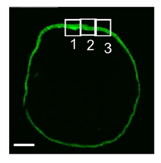
Areas of scanning.
A cross-section of a 10hpf zebrafish Tg(-1.8gsc:GFP)ml1 embryo, labeled with anti-acetylated Tubulin and anti-GFP antibodies (both green). The scale bar is 100 μm. To assess ciliary localization of Smo at different dorso-ventral positions, we scanned three 90 μm areas of each cross-section, as shown. We also examined Smo localization on the ventral side, 180° from the dorsal midline.
Acknowledgments
This image is the copyrighted work of the attributed author or publisher, and
ZFIN has permission only to display this image to its users.
Additional permissions should be obtained from the applicable author or publisher of the image.
Full text @ Curr. Biol.