
IMAGE
Fig. S6
- ID
- ZDB-IMAGE-090710-64
- Genes
- Publication
- Yabe et al., 2009 - The maternal-effect gene cellular island encodes aurora B kinase and is essential for furrow formation in the early zebrafish embryo
- All Figures
- Figures for Yabe et al., 2009
Image
|
Figure Caption
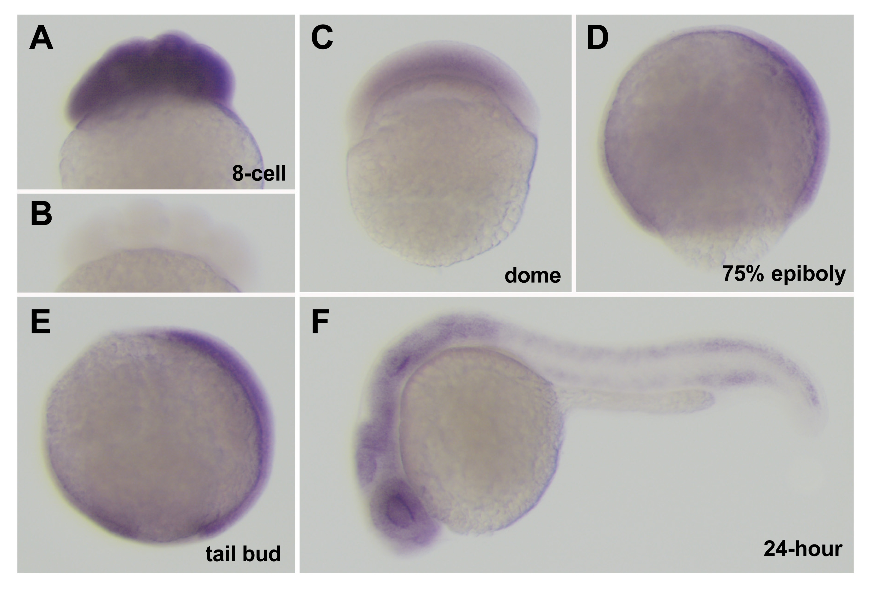
Fig. S6 Expression of cei/aurB mRNA during embryogenesis. (A,C–F) Side views of fixed wild-type embryos processed through in situ hybridization using an antisense (A,C–F) and sense (B) cei/aurB probe. Developmental time points are: 8-cell (75 min p.f.), dome (4.3 hours p.f.), 75% epiboly (8 hours p.f.), tail bud (10 hours p.f.), 24-hour (24 hours p.f.).
Figure Data
Acknowledgments
This image is the copyrighted work of the attributed author or publisher, and
ZFIN has permission only to display this image to its users.
Additional permissions should be obtained from the applicable author or publisher of the image.
Full text @ PLoS Genet.