
|
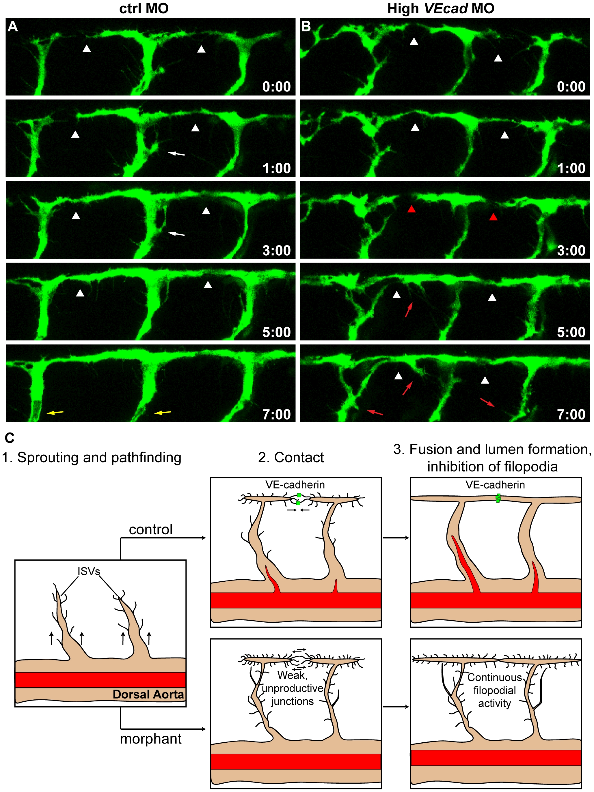
Fig. 7 VE-cadherin is required for correct vascular connection and fusion.
Time lapse analyses of ISVs of ctrl and high VEcadMO injected embryos from 28 to 36 hpf, performed in a Tg(fli1:EGFP) background. Lateral views of the trunk vessels are shown at the indicated time points (see Movies S7 and S8 for full time lapse). In ctrlMO injected embryos (A), dorsal extensions of ISVs that are about to form the DLAV (arrowheads) get stabilized soon after making contact with neighbouring vessels. In addition, the filopodial activity decreases once the connections are formed and stops before lumenization (white arrows). See lumen in ISVs at time point 7:00 (yellow arrows). In contrast, vessels of VE-cadherin severe morphants (B) show defects in establishing stable connections, as they make contact (arrowheads in B) but detach afterwards (red arrowheads). Moreover, the filopodial activity persists in ISVs even once they already appear dorsally interconnected (red arrows). (C) Model of the filopodial sprouting behaviour of control and VE-cadherin severe morphant ISVs.