
|
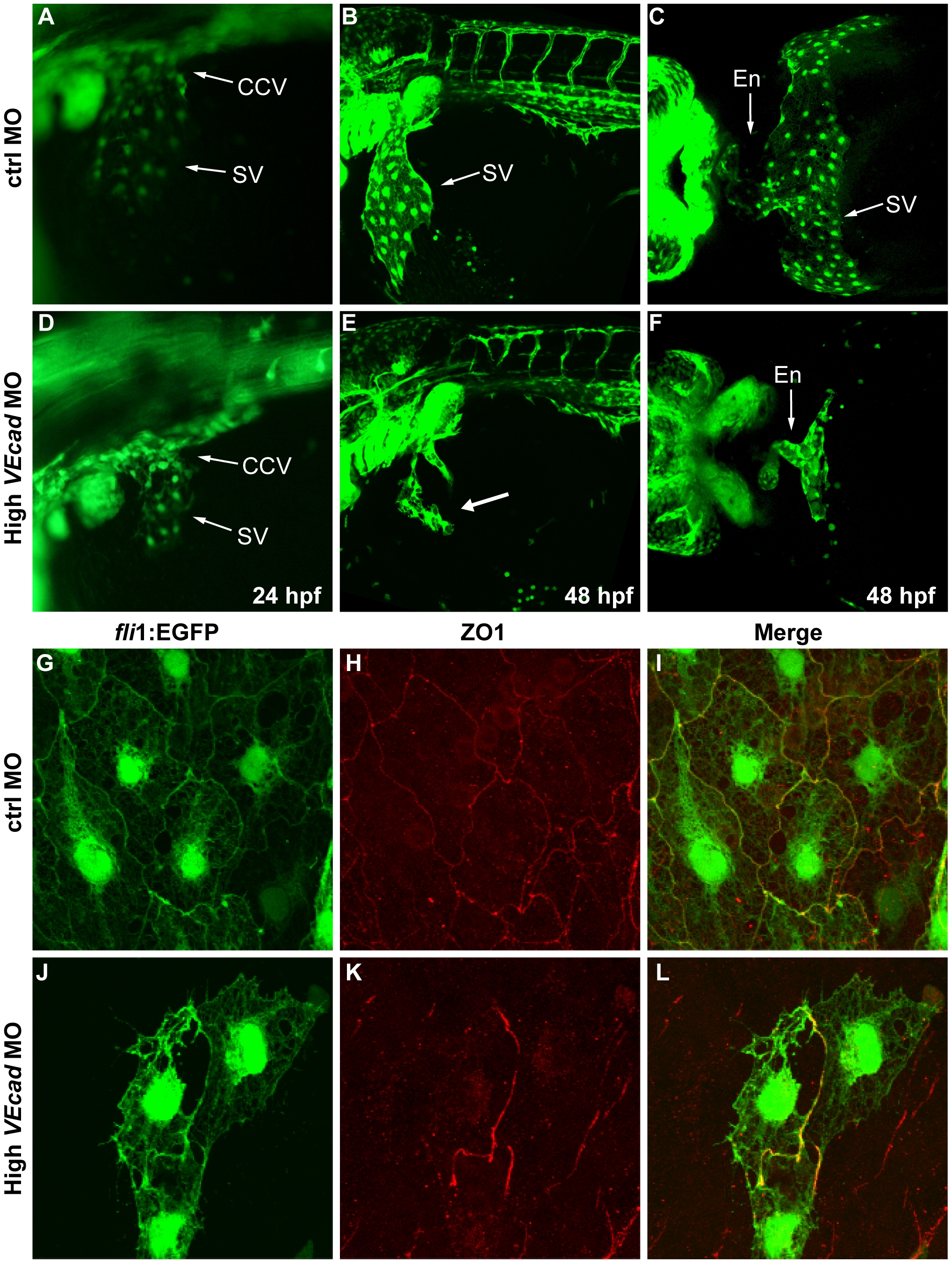
Fig. 5 Depletion of VE-cadherin affects the formation of the sinus venosus.
(A–F) Fluorescence images of fli1:EGFP embryos at the level of the common cardinal vein (CCV) and sinus venosus (SV). At 24 hpf a number of endothelial cells expand out of the CCV to form the SV both present in control embryos (A). In VE-cadherin severe morphant embryos (High VEcadMO) a rudiment of SV appears at 24 hpf (D). At 48 hpf the SV is fully formed in control embryos (B) while in high VEcadMO only few endothelial cells are present (arrow in panel E). Ventral views of fixed embryos allow the visualization of the endocardium (En) connected to a fully formed SV in control embryos (C). In contrast, high VEcadMO do have a tubular endocardium (En in panel F) but the SV is not formed. The absence of this structure impairs the connection of the CCV to the heart tube and the closure of the circulatory loop. (G–L) Immunofluorescence analysis by confocal microscopy of endothelial cells from the SV at 48 hpf. Fli1:EGFP positive endothelial cells visualized in G and J present intercellular junctions, marked by antibody staining against ZO1 (H, K). The few endothelial cells present in this SV region in high VEcadMO injected embryos show an aberrant morphology, but form some junctions at the sites of contact.