
|
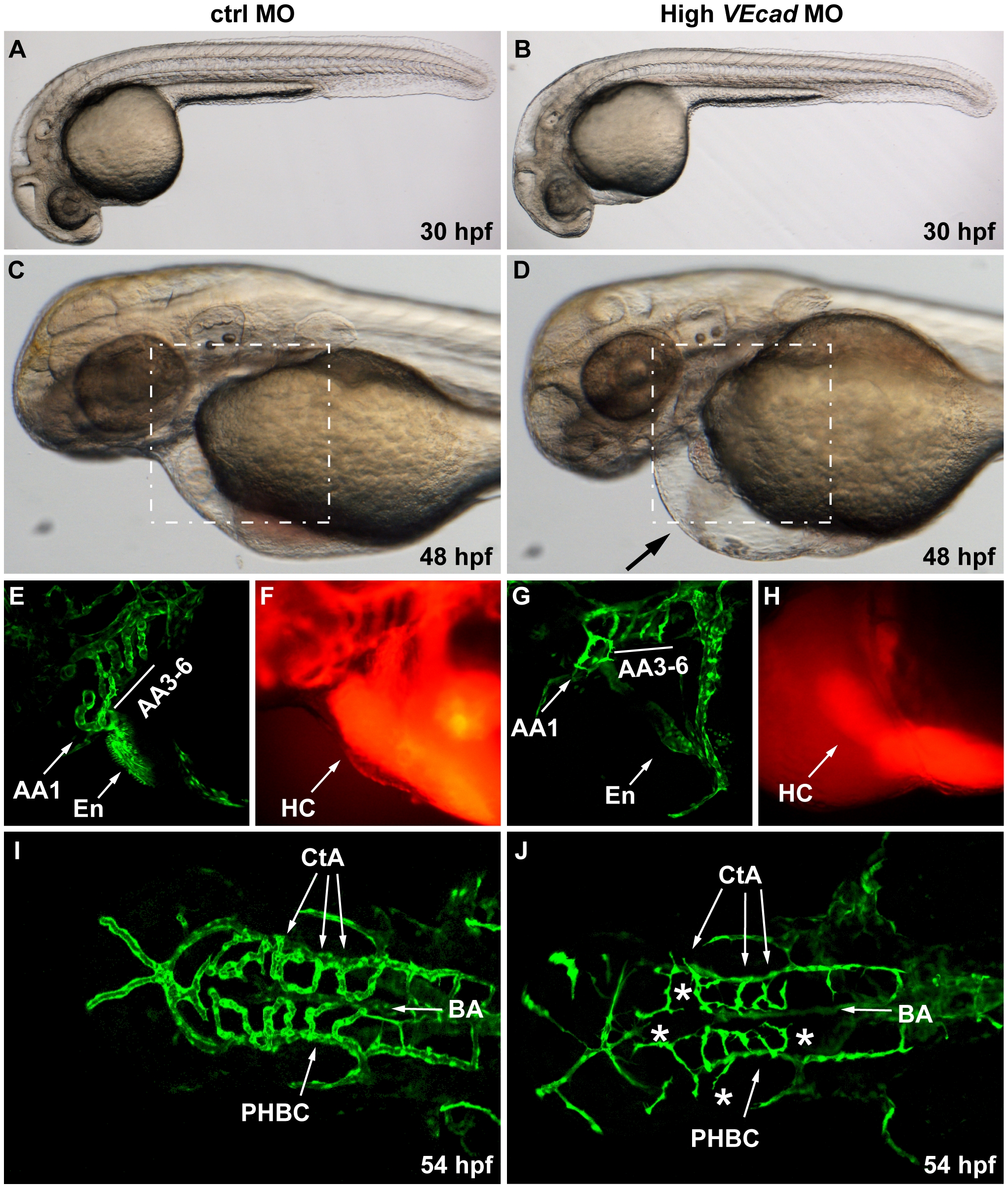
Fig. 4 Complete VE-cadherin abrogation impairs the formation of a functional vascular network.
(A–D) Bright field images of embryos injected with 16 ng of ctrlMO and VE-cadherin morpholino (High VEcadMO). VE-cadherin severe morphants present a normal morphological development at 30 hpf (B) but blood circulation is not established (see movies in supplementary material). At 48 hpf lack of circulation persists in this morphants with severe phenotype, and pericardial edema is observed (arrow in panel D). (E–H) Views of the endocardial and aortic arch region (highlighted box in C and D) at 52 hpf. Endocardium (En) and aortic arch vessels (AA) are visualized using a flk1:EGFP background (E, G) while microangiograms show interconnected lumenized vessels filled with rhodamine-dextan (F). VE-cadherin severe morphants form endocardium (En in panel G) that is not openly connected to the vasculature, neither to the aortic arch 1 (AA1) nor to the CCV, and retains the microinjected dextran in the heart chambers (HC). Rudiments of AAs without lumen are present in VE-cadherin morphants (G). (I, J) Dorsal views of Tg(flk1:EGFP) embryos at 54 hpf. In embryos injected with high dose of VE-cadherin morpholino (J) cranial vessels are apparently in place but central arteries (CtA) fail to form the connection to the basilar artery (BA), do not correctly lumenize and small sprouting vessels are present (asterisks).