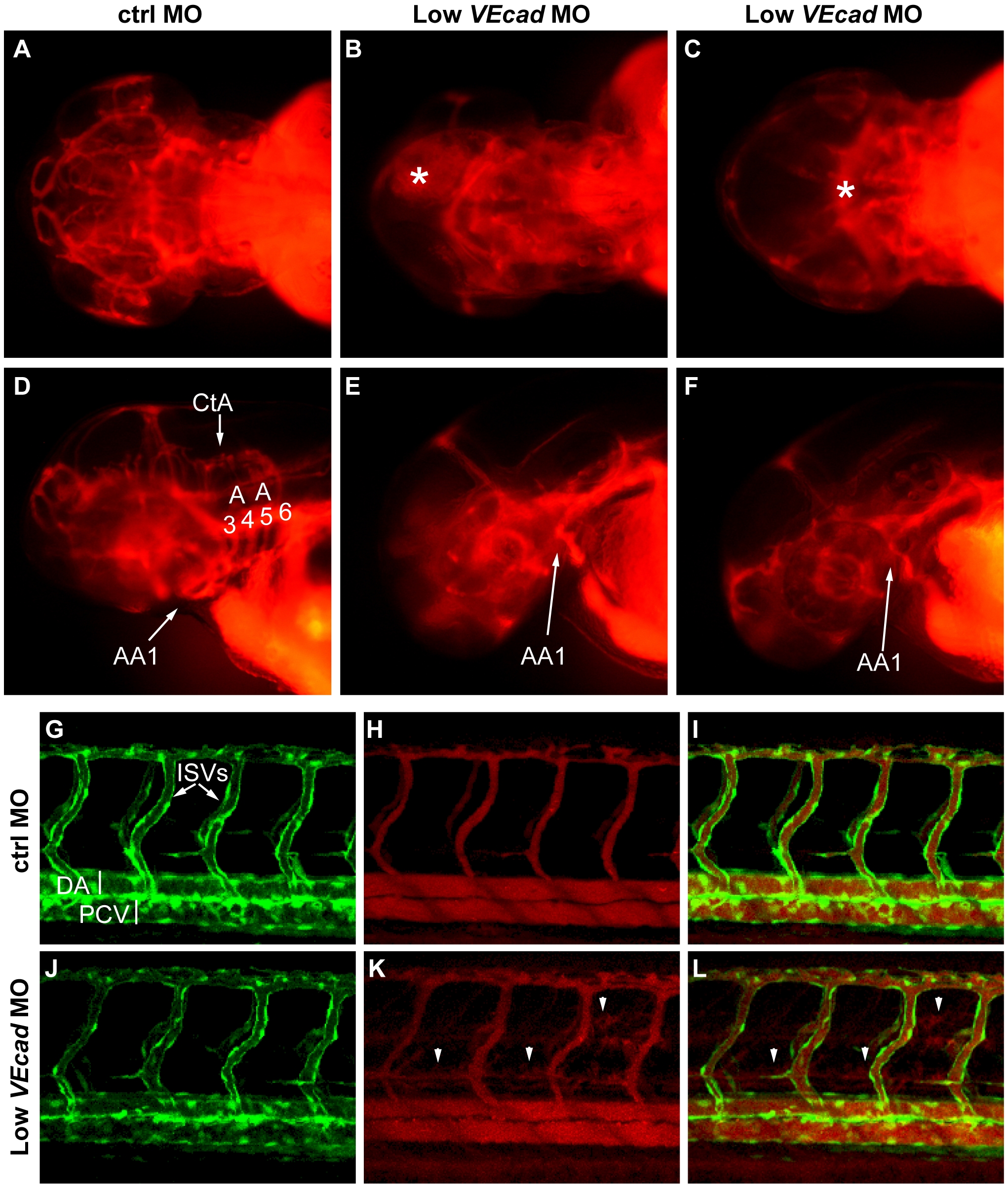
Fig. 3 Partial VE-cadherin knockdown affects cranial and trunk vessel integrity.
(A–F) Microangiographies performed at 52 hpf. (A–C) Dorsal views, (D–F) lateral views of the embryos above, anterior to the left. Control embryos (A, D) have a functional and complex cranial vascular network with lumenized aortic arches (AA) and central arteries (CtA). Microangiograms of embryos injected with low dose (0.8 ng) of VE-cadherin morpholino that developed hemorrhages (Low VEcadMO) show defects in head perfusion. B, E and C, F correspond to two different embryos with the same treatment. Extravasation of the injected dextran is observed at the site of the hemorrhages (asterisk in B and C). In addition, only the first aortic arch (AA1) is fully formed in the mild morphants but aortic arches 3 through 6 do not exhibit circulation (see panels E, F). (G–L) Microangiographies performed in Tg(flk1:EGFP) embryos at 54 hpf. Lateral views of trunk vessels are shown. (G, J) EGFP positive vessels, (H, K) microangiograms performed with rhodamine-dextran, and merge images (I, L). In control embryos (G–I) intersegmental vessels (ISVs), dorsal aorta (DA) and posterior cardinal vein (PCV) are fully lumenized and the injected dextran remains inside this primary vascular network. In some VE-cadherin mild morphants (Low VEcadMO) these vessels are apparently normal (J) but leakage is observed in the somites and regions surrounding the ISVs (arrowheads in panels K,L).

